Review
. 2018 Mar 22;10(4):142.
doi: 10.3390/v10040142.

Rapid Viral Diagnosis of Orthopoxviruses by Electron Microscopy: Optional or a Must?

Affiliations
Review

Rapid Viral Diagnosis of Orthopoxviruses by Electron Microscopy: Optional or a Must?

Hans R Gelderblom et al. Viruses. .

Abstract

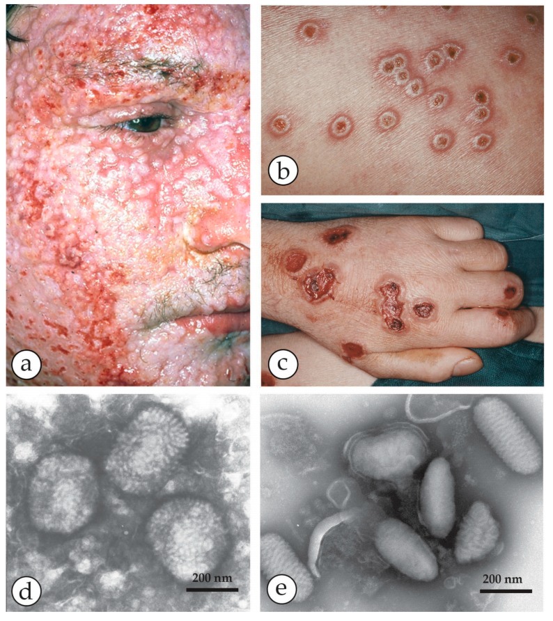
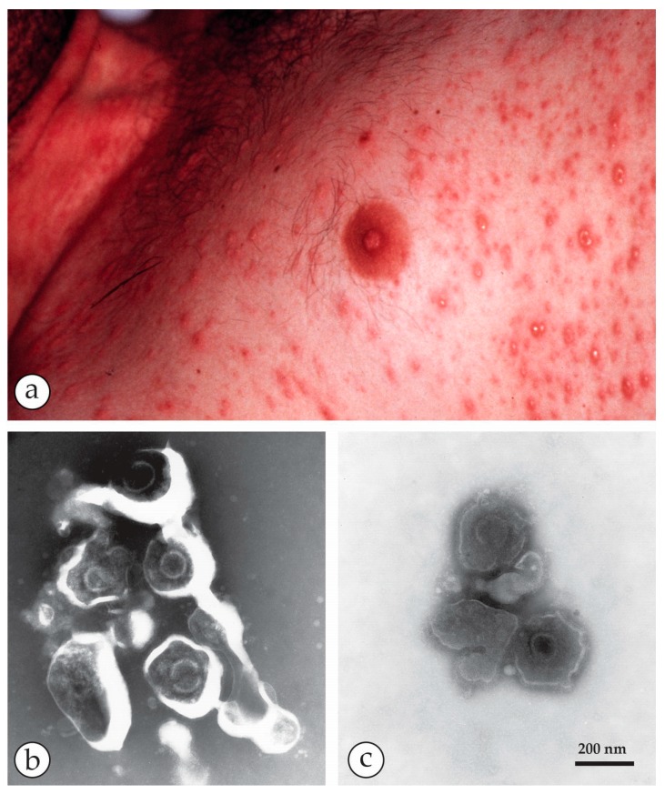
Diagnostic electron microscopy (DEM) was an essential component of viral diagnosis until the development of highly sensitive nucleic acid amplification techniques (NAT). The simple negative staining technique of DEM was applied widely to smallpox diagnosis until the world-wide eradication of the human-specific pathogen in 1980. Since then, the threat of smallpox re-emerging through laboratory escape, molecular manipulation, synthetic biology or bioterrorism has not totally disappeared and would be a major problem in an unvaccinated population. Other animal poxviruses may also emerge as human pathogens. With its rapid results (only a few minutes after arrival of the specimen), no requirement for specific reagents and its "open view", DEM remains an important component of virus diagnosis, particularly because it can easily and reliably distinguish smallpox virus or any other member of the orthopoxvirus (OPV) genus from parapoxviruses (PPV) and the far more common and less serious herpesviruses (herpes simplex and varicella zoster). Preparation, enrichment, examination, internal standards and suitable organisations are discussed to make clear its continuing value as a diagnostic technique.

Keywords: VZV); diagnostic electron microscopy (DEM); febrile vesicular rashes; herpesviruses (HSV; negative staining; orthopoxviruses (OPV); parapoxviruses (PPV); rapid viral diagnosis; skin lesions.


Conflict of interest statement

The authors declare no conflict of interests. The conclusions drawn are those of the authors and do not necessarily represent the views of our institutions.

Figures

Figure 1
Figure 1
Last cases of endemic smallpox in Europe: (a,b) Smallpox patient from the 1962 outbreak in South Wales. (a) At the acute stage, with marked facial oedema, and (b) after recovery. He was 17 at the time, but looked middle-aged in the first photo. (c) The hand of one of the cases showing deep-seated, compact vesicles, often umbilicated and mostly at the same developmental stage. (d) Negative staining (NS) DEM of vesicle fluid with PTA (potassium phosphotungstic acid) revealed brick-shaped particles of the proper OPV size. (e,f) Thin section (TS) TEM (Transmission Electron Microscopy) and DEM of the last smallpox case in Germany [30]. (e) Virus was isolated on the chorio-allantoic membrane (CAM) and showed in ultrathin sections different cuts through fully assembled OPV. For more detail see Figure 14a,b. (f) direct DEM of vesicle fluid showed an abundance of typical OPV particles. (f) with kind permission of Thieme Verlag, Stuttgart, Germany. Bars (df) = 200 nm.
Figure 2
Figure 2
Variolation versus vaccination: (a) Variolation, the inoculation of small amounts of live variola lesion fluids was widely used as a protective, though highly risky measure, before Edward Jenner in 1796 established the much less risky vaccination using material from a cowpox lesion [31]. Plate from Fenner et al.: Smallpox and its Eradication [1] with kind permission of WHO, Geneva. (b) A routine primary vaccination lesion in a one-year old, containing little vesicle fluid. Photo taken after 1 week. (c) Vaccinia virus after NS showing all morphological criteria: size, shape and surface details of OPV. The origin of present day vaccines strains is heterogeneous. For details see [1,16,24,32].
Figure 3
Figure 3
A fatal case of a cowpox zoonosis: (ac) A zoonotic CPXV infection in an 18 year old immuno-compromised man. He was under massive steroid therapy for an allergy. The patient took care of a stray cat and developed fever and a generalized rash 10 days later. Vesicles developed into hemorrhagic pustules (b) with a tendency to fuse into larger ulcers (c). The patient died with circulatory collapse. (d) NS-DEM of specimens with PTA revealed brick-shaped particles with short surface threats typical of OPV. (ad) reprinted from [33], with kind permission of Drs. Anna M. Eis-Hübinger and Bernhard Pfeiff and Springer Nature. (e) For comparison Parapoxviruses (PPV) after NS: smaller than OPV, ovoid and surrounded by long parallel spiral surface threads.
Figure 4
Figure 4
Peculiarities of CPXV infections: wide host range and specific inclusion bodies. (a) Lesions on the inner surface of a trunk of a deceased elephant, sliced longitudinally showing multiple ulcerous lesions on the mucous membranes [34]. Image courtesy Dr. Gudrun Wibbelt, Berlin. (b) After inoculation from the elephant´s ulcers in diagnostic cell cultures, well-circumscribed inclusion bodies developed consisting of a moderately dense matrix that included numerous mature virions. These are type A eosinophilic inclusions as seen in light microscopical histology and called Downie or Marchal bodies. They are typical of CPXV but found with a few other poxvirus genera [35]. (c) OPV are assembled in large cytoplasmic “factories” but these are basophilic (type B inclusions or Guarnieri bodies) in light microcopy and less well circumscribed.
Figure 5
Figure 5
Human monkeypox in the USA in 2003 [36,37]. (a) The 2003 multi-state outbreak of human monkeypox in the Mid-West of the USA was initiated by an imported Gambian giant rat carrying MPXV. The rat infected prairie dogs kept as pets and the virus was transmitted to humans as zoonotic infections. The patients developed mild fever, lymphadenopathy and localized lesions where their pet animals had bitten or scratched them. (b) Vesicles developed into deep-seated pustules and slightly hemorrhagic ulcers that dried later. (c) The etiology remained unclear for 10 days until DEM was used, revealing typical OPV particles. Based on this orientation the final diagnosis of MPXV was confirmed at CDC [37]. Reprinted with kind permission of New England Journal Medicine.
Figure 6
Figure 6
Outbreaks of human MPXV infections in Africa: (a) A boy from Democratic Republic of Congo with a generalized MPXV zoonosis. The vesicular lesions appear solid and are nearly all at the same developmental state (Photo courtesy of Mark Szczeniowski, WHO). After smallpox had been eradicated, human MPXV raised major concerns as an emerging zoonosis. Thirty years after the end of smallpox vaccination, the rate of this zoonosis increased 20-fold, in part due to increased contact with infected bushmeat [25]. Laboratory diagnosis showed, however, MPXV being responsible for only half the suspected “zoonoses”, the other half being caused by VZV [38,39]. DEM run in parallel at the Bernhard-Nocht-Institute in Hamburg by Christel Schmetz and at the Robert Koch Institute in Berlin confirmed the diagnosis. The samples for DEM had been kindly given by Prof. Hermann Meyer, Munich as aldehyde-inactivated samples and less than 5 µL each. Nevertheless, the particles shown in (b,c) have clearly typical herpesvirus and OPV-morphology respectively. Bars = 200 nm.
Figure 7
Figure 7
DEM of an Orf-zoonosis: (a) Index finger with a confluent haemorrhagic ulcer of a farmer who had handled his Orf-infected sheep [40]. (b) The “roof” of a lesion was homogenized. After NS with PTA ovoid particles are readily detected among some cellular detritus. The number of particles observed is consistent with a concentration of 107–8 particles per mL in the original specimen. (c) Same case: The five virions shown at higher magnification are typical members of the PPV genus: they are smaller than OPV or MCV, differ by having an ovoid shape and present long, parallel running apparently spiral surface threads, Sample and clinical image courtesy of Prof. Klaus Eisendle, Bolzano). (b,c) reproduced from [40] with kind permission of Elsevier.
Figure 8
Figure 8
DEM of another human PPV infection (sample courtesy of Prof. Friedrich A. Bahmer, Bremen): (a) TS-DEM of a biopsy of a papule showing numerous particulate objects: on the right, PPV particles can be seen while the numerous electron-dense structures on the left are melanosomes–normal skin constituents Bar in (a) = 1 μm. In the middle are desmosomes connecting cells in the prickle cell layer. (b) TS-DEM at an intermediate magnification reveals the oval shapes typical of PPV seen in a damaged cell. (c) PPV particles are seen also by NS-DEM after grinding parts of the biopsy in distilled water, followed by low speed clarifying centrifugation. Amongst some detritus and a long collagen fibre, the ovoid PPV particles show long parallel surface threads. Bars in (b,c) = 200 nm.
Figure 9
Figure 9
Molluscipox virus lesions: (a) Face of a young boy from Tanzania presenting numerous solid, yellow-whitish skin nodules typical of Molluscum contagiosum (photo courtesy of Prof. Constantin Orfanos, Berlin). (b,c): Ultrathin section TEM (TS-TEM) of a biopsy of a MCV papule. While the low power micrograph (b) shows the abundance of virus particles in the lobulated compartments of the nodule, the higher magnification in (c) reveals different orientations of MCV virions. By TS- and by NS-DEM, MCV are indistinguishable from OPV. (d) and (e): NS-DEM of the contents of MC papules: (d) After grinding a biopsy, six virions are shown which by size and shape and by the irregular surface structure closely resemble OPV. The virions are seen amid a meshwork of cell remnants and collagen fibres, the latter with their typical repeat pattern of 67 nm. (e) DEM without taking a surgical biopsy: the waxy content of a MC nodule was extruded by gentle squeezing using forceps. After dilution in distilled water, numerous brick-shaped particles, free from cellular contaminants were seen. DEM on many other skin lesions can be performed without using surgery [44]. Bars in (ce) = 200 nm.
Figure 10
Figure 10
Chickenpox, a common, mild febrile rash in childhood. (a) Clinical chickenpox in a five-year old child. The lesions are mostly on the trunk. (b) Close-up of vesicles on the arm. They are more superficial compared with the deep-seated OPV or specifically those of smallpox. (c) NS-DEM of the clean vesicle fluid shows three herpesviruses. The labile virions are penetrated by the UAc stain and reveal ruptured envelopes containing the hexagonal central 110 nm capsids.
Figure 11
Figure 11
Shingles: (a) A case of shingles, a reactivated VZV infection, with its typical unilateral distribution of the vesicles, usually confined to a single dermatome. (b) Fluid collected from a vesicle later in the illness shows an aggregate of herpesvirus particles, penetrated by the PTA-stain, and mixed with some cell detritus. Combining DEM results with the clinical appearance and distribution of the lesions confirms the diagnosis of a reactivated VZV infection.
Figure 12
Figure 12
Herpes simplex eruption. (a) Severe herpes simplex eruptions on the lips and chin of a young woman. (b) Typical herpes virus particles seen after PTA NS of vesicle fluid. The labile envelopes are stain-penetrated and reveal the similarly stain-penetrated capsids. In the thinner stain, the upper virion also shows glycoprotein spikes in the periphery of its envelope.
Figure 13
Figure 13
A comparison of OPV, PPV and herpesviruses after NS- (left) and TS- (right) preparation and DEM: (a) NS-DEM using UAc of a suspension of ectromelia OPV showing “brick-shaped” particles, 250 × 350 nm in size, with an irregular pattern of short, 10–15 nm surface protrusions. (b) TS-DEM of OPV from a diagnostic CAM from the last smallpox case in Germany [30]. Within the sections through the virions, inner components, the dumbbell-shaped core and lateral bodies can be seen. (c) NS-DEM of PPV observed in a diagnostic biopsy after grinding and NS with UAc. The ovoid virion is surrounded by parallel-running surface threads and lying beside a thick thread of collagen with its typical 67 nm-periodicity. The case was diagnosed later as a zoonotic infection of a butcher by bovine papular stomatitis virus. (d) TS-TEM of PPV observed in the biopsy from the same case as (c). In longitudinal section planes, PPV appear slimmer than OPV and they also lack the prominent lateral bodies of OPV. (e) NS of herpes virus capsids, showing occasionally a roughly hexagonal outline. The electron-translucent portions inside the core contain nucleoproteins associated with the viral DNA. (f) TS-TEM of a diagnostic culture showing two virions of equine herpesvirus (EHV-1). The envelope is studded with fuzzy surface projections. Underneath the lipid bilayer the ill-defined tegument and the slightly angular cores are seen. The latter contains the electron-dense viral genome.
Figure 14
Figure 14
NS, the most rapid technique in diagnostic virology. It is done using a non-wetting surface, such as a sheet of ParafilmTM, and comprises the following steps: adsorption, washing and staining. A hydrophilic EM support grid is placed on a droplet of the diagnostic suspension (left) and after 30 s of adsorption is transferred quickly onto a series of droplets of distilled water to remove interfering salts, and then onto a droplet of “stain”. The “stains” used are electron-dense solutions: 0.5 to 2.0 percent of a heavy metal salt, such as PTA, routinely buffered to pH 7.2 (may be used between pH 5 to pH 10), UAc, unbuffered at a pH around 4.2, or other stains can be used. During a short, 5–10 s staining step, the stain does not react with the chemical moieties of the biologicals on the grid, i.e., there is no “positive staining” effect as there is in thin section-TEM. This rapid process results in “negative contrast”, as the transparent biological structures of the sample, after drying, are closely surrounded by the electron-dense stain. Reprinted from [45] with the kind permission of Springer Nature.
Figure 15
Figure 15
An unexpected result: (a) A presumed syphilitic lesion on the tip of the tongue of a young mother. When dark field light microscopy repeatedly failed to reveal Treponema pallida, doubts about the cause arose, and the patient was sent to hg for DEM to look for a viral cause. (b) Grids, touched onto the ulcerous surface, were stained with 2% PTA, pH 8.5, and subsequently inactivated by formaldehyde steam. Brick-shaped structures, 400 × 250 nm in size, were seen, most of them disintegrated and flattened. The internal “triple coil” (the DNA-containing inner body) is typical of OPV [92] (c) In the even stain, some surface detail and the overall size and shape are revealed. (b,c) are at the same magnification and reproduced from [93] with kind permission of Springer Nature.
Figure 16
Figure 16
“An unconscious, unknown patient”: (a) Thin-walled blisters, differing in size and development on the thorax of an unconscious febrile patient. To collect fluid for rapid DEM, vesicles were opened and EM grids touched onto the vesicle´s base. (b) NS-DEM with 2% PTA revealed an abundance of herpesviruses. In the relatively ‘deep’ PTA-stain, the virions are morphologically degraded, i.e., the viral envelopes are broken open and some cores damaged by osmotic damage and shrinkage of the stain as it dries. (c) Three herpesvirus particles seen in a flat and even PTA-stain. Viral envelopes and cores retain the PTA stain as in (b). The measured diameter of the cores and the appearance of the envelope remnants allow a safe diagnosis: ‘particles of the order Herpesvirales.’ (b,c) are at the same magnification and reproduced from [73] (with kind permission of New Microbiologica).

Similar articles

Cited by

References

    1. Fenner F., Henderson D.A., Arita I., Jezek L., Ladnyi I.D. Smallpox and Its Eradication. World Health Organization; Geneva, Switzerland: 1988.
    1. Essbauer S., Pfeffer M., Meyer H. Zoonotic poxviruses. Vet. Microbiol. 2010;140:229–236. doi: 10.1016/j.vetmic.2009.08.026. - DOI - PMC - PubMed
    1. Morens D.M., Folkers G.K., Fauci A.S. The challenge of emerging and re-emerging infectious diseases. Nature. 2004;430:242–249. doi: 10.1038/nature02759. - DOI - PMC - PubMed
    1. Shchelkunov S.N. An increasing danger of zoonotic orthopoxvirus infections. PLoS Pathog. 2013;9:e1003756. doi: 10.1371/journal.ppat.1003756. - DOI - PMC - PubMed
    1. Olson V., Shchelkunov S. Are we prepared in case of a possible smallpox-like disease emergence? Viruses. 2017;9:242. doi: 10.3390/v9090242. - DOI - PMC - PubMed

MeSH terms

LinkOut - more resources