Review
. 2018 Mar 22;19(4):942.
doi: 10.3390/ijms19040942.

Role of Inflammation in Diabetic Retinopathy

Affiliations
Review

Role of Inflammation in Diabetic Retinopathy

Anne Rübsam et al. Int J Mol Sci. .

Abstract

Diabetic retinopathy is a common complication of diabetes and remains the leading cause of blindness among the working-age population. For decades, diabetic retinopathy was considered only a microvascular complication, but the retinal microvasculature is intimately associated with and governed by neurons and glia, which are affected even prior to clinically detectable vascular lesions. While progress has been made to improve the vascular alterations, there is still no treatment to counteract the early neuro-glial perturbations in diabetic retinopathy. Diabetes is a complex metabolic disorder, characterized by chronic hyperglycemia along with dyslipidemia, hypoinsulinemia and hypertension. Increasing evidence points to inflammation as one key player in diabetes-associated retinal perturbations, however, the exact underlying molecular mechanisms are not yet fully understood. Interlinked molecular pathways, such as oxidative stress, formation of advanced glycation end-products and increased expression of vascular endothelial growth factor have received a lot of attention as they all contribute to the inflammatory response. In the current review, we focus on the involvement of inflammation in the pathophysiology of diabetic retinopathy with special emphasis on the functional relationships between glial cells and neurons. Finally, we summarize recent advances using novel targets to inhibit inflammation in diabetic retinopathy.

Keywords: Müller glial cells; astrocytes; crystallins; diabetic retinopathy; inflammation; microglia; neurodegeneration; neuroprotection; pathophysiology.


Conflict of interest statement

The authors declare no conflict of interest.

Figures
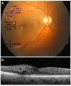
Figure 1
Figure 1
Clinical features of diabetic retinopathy and causative pro-inflammatory chemokines. (A) A fundus photograph shows the right eye of a 57-year-old man with 20/80 visual acuity and signs of severe non-proliferative diabetic retinopathy with non-significant macular edema (the region of macular edema is indicated by the bracket). Vascular pathologies are depicted in blue, whereas neurodegenerative features are black. Notable features include dot blot hemorrhages, DH; hard exudates, HE; cotton-wool spots, CWS. Each are associated with the upregulation of certain chemokines; (B) Optical coherence tomography with a horizontal scan through the central fovea reveals moderate thickening and edema of the macula with cysts; (C) Ang: Angiopoietin; IL: interleukin; TNF: Tumor Necrosis Factor; NO: nitric oxide; MCP: Monocyte Chemoattractant Protein.
Figure 2
Figure 2
Schematic representation of pathogenic mechanisms leading to sight-threatening endpoints of diabetic retinopathy (DR): proliferative DR (PDR) and diabetic macular edema (DME). Metabolic alterations are first sensed by glial cells resulting in glial dysfunction, which induces inflammation, aberrant signaling of trophic factors and metabolic dysregulation all leading to neuronal apoptosis. Neurodegeneration also participate in blood–retinal barrier (BRB) breakdown, vasoregression and consecutive hypoxia, the main features of early microvascular abnormalities and the end stage neovascularization. According to the vascular lesions DR is staged by ophthalmoscopy in several stages of severity. AGE, advanced glycation end-products; PKC, protein kinase C; RAS, renin–angiotensin system, ROS, reactive oxygen species; NADPH, Nicotinamide adenine dinucleotide phosphate; mETC, mitochondrial electron transport chain; NPDR, non-proliferative DR.
Figure 3
Figure 3
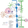
Schematic of a mammalian retina depicting the interactions between glia, neurons and endothelial cells with special regard to the inflammatory chemokines involved. Blood vessels and endothelial cells in pink (9), leukocytes in purple (5), macroglial cells in green (7,8), microglia in brown (2,3) and neurons in blue (6ac). Scheme showing kalium homeostasis, glutamate metabolism and the secretion of trophic factors, chemokines and interleukins. AGEs, advanced glycation end products.

References

    1. Klein B.E. Overview of epidemiologic studies of diabetic retinopathy. Ophthalmic Epidemiol. 2007;14:179–183. doi: 10.1080/09286580701396720. - DOI - PubMed
    1. Kempen J.H., O’Colmain B.J., Leske M.C., Haffner S.M., Klein R., Moss S.E., Taylor H.R., Hamman R.F., Eye Diseases Prevalence Research Group The prevalence of diabetic retinopathy among adults in the United States. Arch. Ophthalmol. 2004;122:552–563. - PubMed
    1. Zhang X., Saaddine J.B., Chou C.F., Cotch M.F., Cheng Y.J., Geiss L.S., Gregg E.W., Albright A.L., Klein B.E., Klein R. Prevalence of diabetic retinopathy in the United States, 2005–2008. JAMA. 2010;304:649–656. doi: 10.1001/jama.2010.1111. - DOI - PMC - PubMed
    1. AAO PPP Retina/Vitreous Panel, Hoskins Center for Quality Eye Care. [(accessed on 30 October 2017)];Diabetic Retinopathy Ppp—Updated. 2016 Available online: http://www.aao.org/preferred-practice-pattern/diabetic-retinopathy-ppp--....
    1. Frey T., Antonetti D.A. Alterations to the blood-retinal barrier in diabetes: Cytokines and reactive oxygen species. Antioxid. Redox Signal. 2011;15:1271–1284. doi: 10.1089/ars.2011.3906. - DOI - PubMed