. 2018 May;39(5):2081-2090.
doi: 10.3892/or.2018.6311. Epub 2018 Mar 14.

CAFs enhance paclitaxel resistance by inducing EMT through the IL‑6/JAK2/STAT3 pathway

Affiliations

CAFs enhance paclitaxel resistance by inducing EMT through the IL‑6/JAK2/STAT3 pathway

Linlin Wang et al. Oncol Rep. 2018 May.

Abstract

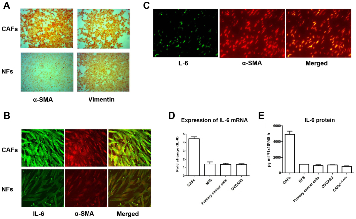
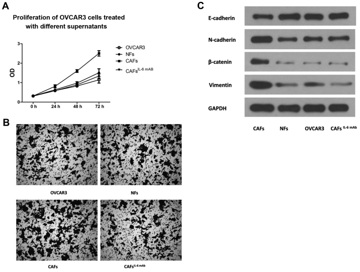
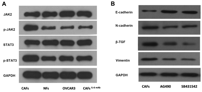
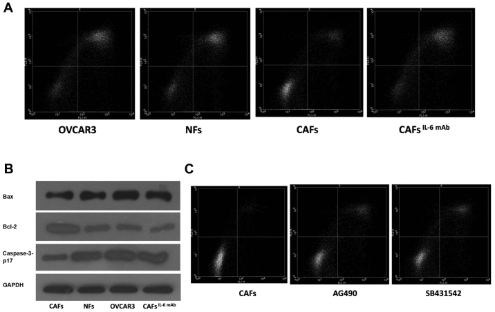
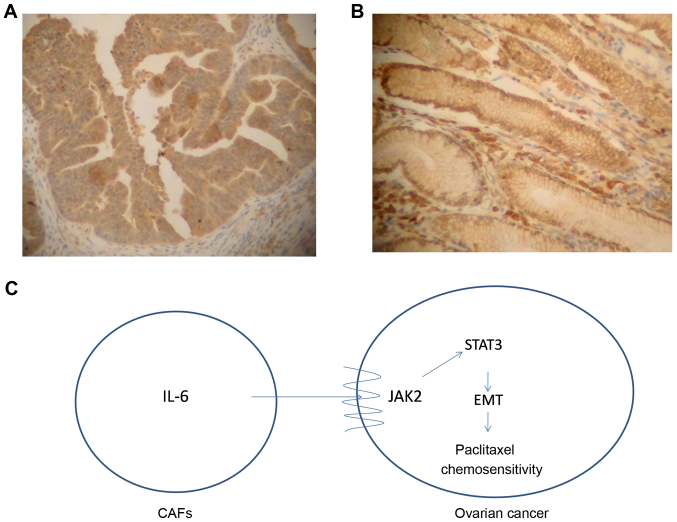
Carcinoma‑associated fibroblasts (CAFs) are the major components of mesenchymal cells in the inflammatory tumor microenvironment. They are involved in epithelial‑mesenchymal transition (EMT) and chemotherapy resistance by directly contacting with cancer cells or secretory cytokines. In the present study, we examined the role of CAFs in the induction of EMT in ovarian cancer. Primary ovarian cancer cells, CAFs and normal fibroblasts (NFs) were isolated from fresh cancer tissue and cultured for immunohistochemistry studies. Enzyme‑linked immunosorbent assay (ELISA) was used to detect the expression of IL‑6 in the culture supernatants of these cells. The expression of IL‑6 at the mRNA level was examined by RT‑PCR. The expression of IL‑6 at the protein level in ovarian cancer tissues was determined using an immunofluorescence assay in both tissue sections and cell lobes. OVCAR3 cells were treated with the culture supernatants collected from CAFs and NFs. IL‑6 monoclonal antibody (mAb) was employed to neutralize IL‑6. The expression of phosphorylated STAT3 was assessed. Changes in EMT, proliferation, invasion and proapoptotic protein expression were also examined. Flow cytometry was performed to detect the changes in apoptosis resistance of OVCAR3 cells. The JAK2/STAT3 pathway‑specific inhibitor AG490 was used to block this pathway and the β‑TGF inhibitor was used to inhibit EMT. The clinical data of patients treated in our hospital were collected between January 1st, 2009 and June 30th, 2013. The expression of interstitial IL‑6 in paraffin‑embedded tissues was detected by immunohistochemistry. The relationship between the expression of interstitial IL‑6 and the treatment response was examined by linear regression and multiple linear regression analyses. We found that CAFs were the main source of IL‑6 in ovarian cancer tissue. CAFs promoted the phosphorylation of STAT3 in ovarian cancer and enhanced the proliferation, invasion and EMT. Enhanced EMT may lead to apoptosis resistance, inhibitory expression of pro‑apoptotic proteins and paclitaxel resistance. A total of 255 patients were enrolled in this retrospective study. Univariate and multivariate analyses revealed that age, CA125, interstitial IL‑6 expression and cytoreduction satisfaction were closely related to the sensitivity of the TP (docetaxel plus cisplatin or carbopatin) regimen in ovarian cancer (P<0.05). These results demonstrated that CAFs highly secreted IL‑6 and promoted β‑TGF‑mediated EMT in ovarian cancer via the JAK2/STAT3 pathway, leading to inhibited apoptosis and subsequent paclitaxel resistance. Therefore, CAFs may be a new therapeutic target for the treatment of ovarian cancer.


Figures

Figure 1.
Figure 1.
CAFs are the main source of IL-6 secretion. (A) Following the separation and purification of CAFs and NFs, the cells were stained with α-SMA (100-fold) and Vimentin (100-fold). Both CAFs and NFs expressed Vimentin. However, α-SMA was almost not expressed in NFs, while it was overexpressed in CAFs. (B) CAFs and NFs were stained with IL-6 and α-SMA (100-fold). The expression of IL-6 and α-SMA was positive in CAFs. The expression of IL-6 and α-SMA detected in NFs was weak. (C) Immunofluorescence staining of IL-6 (100-fold) and α-SMA (100-fold) was performed on histological sections of EOC. Similarly, these results revealed that IL-6 was mainly expressed in the interstitium and interstitial α-SMA-labelled CAFs. (D) The culture supernatants of CAFs, NFs, primary cancer cells and OVCAR3 cells were extracted to detect the expression of IL-6 by ELISA. The expression of IL-6 in CAFs was significantly higher than that in NFs, primary cancer cells and OVCAR3. (E) Detected by RT-PCR, the expression of IL-6 at the mRNA level in CAFs was significantly higher than that in NFs, primary cancer cells and OVCAR3.
Figure 2.
Figure 2.
CAF-derived IL-6 promotes the proliferation, migration and EMT of ovarian cancer cells. The culture supernatants of CAFs and NFs were collected and applied to OVCAR3 cells. (A) CCK-8 proliferation assay revealed that the supernatant derived from CAFs could significantly enhance the proliferation of OVCAR3 cells. Similarly, the invasive potential of OVCAR3 cells treated with CAF supernatant was significantly higher than that of NF supernatant. However, with the addition of IL-6 mAb, the effect of CAF supernatant on the proliferation and invasion of OVCAR3 cells was significantly reduced. (B) The effect of CAF supernatant on the expression of EMT markers was significantly attenuated by the addition of IL-6 mAb. There was no significant change in EMT markers after treatment with NFs supernatant. (C) Western blot analysis revealed that the OVCAR3 cells had significantly increased expression of the interstitial markers N-cadherin and Vimentin, and decreased expression of the epithelium marker E-cadherin, suggesting that the EMT of OVCAR3 cells was significantly enhanced.
Figure 3.
Figure 3.
The JAK2/STAT3 pathway is required for regulating EMT in ovarian cancer cells induced by IL-6. (A) The culture supernatants of CAFs and NFs were applied to OVCAR3 cells. The phosphorylation levels of JAK2 and STAT3 in OVCAR3 cells treated with CAF supernatant were significantly higher than those in cells treated with NF supernatant. After the addition of IL-6 mAb, the phosphorylation levels of JAK2 and STAT3 were decreased. (B) After the JAK2/STAT3-signaling-pathway-specific inhibitor AG490 was added, the expression of the interstitial markers N-cadherin and Vimentin was decreased and the expression of the epithelium marker E-cadherin was increased. These results indicated that CAF-derived IL-6 could mediate EMT in OVCAR3 cells via the JAK2/STAT3 pathway.
Figure 4.
Figure 4.
CAF-derived IL-6 enhances paclitaxel resistance of ovarian cancer cells through cellular EMT. (A) The culture supernatants of CAFs and NFs were applied to OVCAR3 cells. The number of apoptotic cells was decreased in OVCAR3 cells treated with CAF supernatant. After the addition of IL-6 mAb, paclitaxel resistance was reduced and paclitaxel-induced apoptosis was promoted. (B) The expression of pro-apoptotic protein Bax and caspase-3-p17 was decreased, and the expression of apoptosis-suppressing protein Bcl-2 was enhanced in cells treated with CAF supernatant compared with NF supernatant. (C) The number of apoptotic cells treated with paclitaxel was increased after the addition of SB431542 and AG490.
Figure 5.
Figure 5.
The expression of IL-6 in tumor stroma. (A) Low expression of interstitial IL-6 protein in paraffin-embedded tissues (100-fold). (B) High expression of interstitial IL-6 protein in paraffin-embedded tissues (100-fold). (C) Schematic diagram which demonstrates that CAFs secrete IL-6 and promote β-TGF-mediated EMT in ovarian cancer cells via the JAK2/STAT3 signaling pathway, leading to the proliferation and invasion of ovarian cancer cells, as well as to apoptosis inhibition and paclitaxel resistance.

References

    1. Siegel RL, Miller KD, Jemal A. Cancer statistics, 2017. CA Cancer J Clin. 2017;67:7–30. doi: 10.3322/caac.21387. - DOI - PubMed
    1. Polyak K, Weinberg RA. Transitions between epithelial and mesenchymal states: Acquisition of malignant and stem cell traits. Nat Rev Cancer. 2009;9:265–273. doi: 10.1038/nrc2620. - DOI - PubMed
    1. Thiery JP, Acloque H, Huang RY, Nieto MA. Epithelial-mesenchymal transitions in development and disease. Cell. 2009;139:871–890. doi: 10.1016/j.cell.2009.11.007. - DOI - PubMed
    1. Erez N, Glanz S, Raz Y, Avivi C, Barshack I. Cancer associated fibroblasts express pro-inflammatory factors in human breast and ovarian tumors. Biochem Biophys Res Commun. 2013;437:397–402. doi: 10.1016/j.bbrc.2013.06.089. - DOI - PubMed
    1. Del Prete A, Allavena P, Santoro G, Fumarulo R, Corsi MM, Mantovani A. Molecular pathways in cancer-related inflammation. Biochem Med. 2011;21:264–275. doi: 10.11613/BM.2011.036. - DOI - PubMed

MeSH terms