. 2018 Apr 20;7(4):1116-1130.
doi: 10.1021/acssynbio.8b00018. Epub 2018 Mar 29.

Generation of a Synthetic Human Chromosome with Two Centromeric Domains for Advanced Epigenetic Engineering Studies

Affiliations

Generation of a Synthetic Human Chromosome with Two Centromeric Domains for Advanced Epigenetic Engineering Studies

Elisa Pesenti et al. ACS Synth Biol. .

Abstract

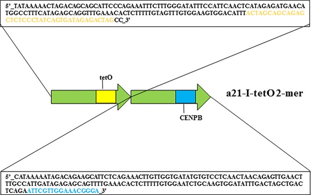
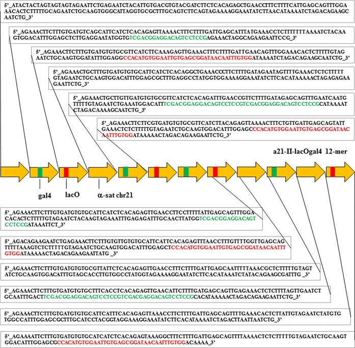
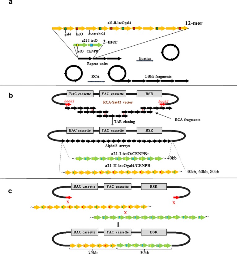
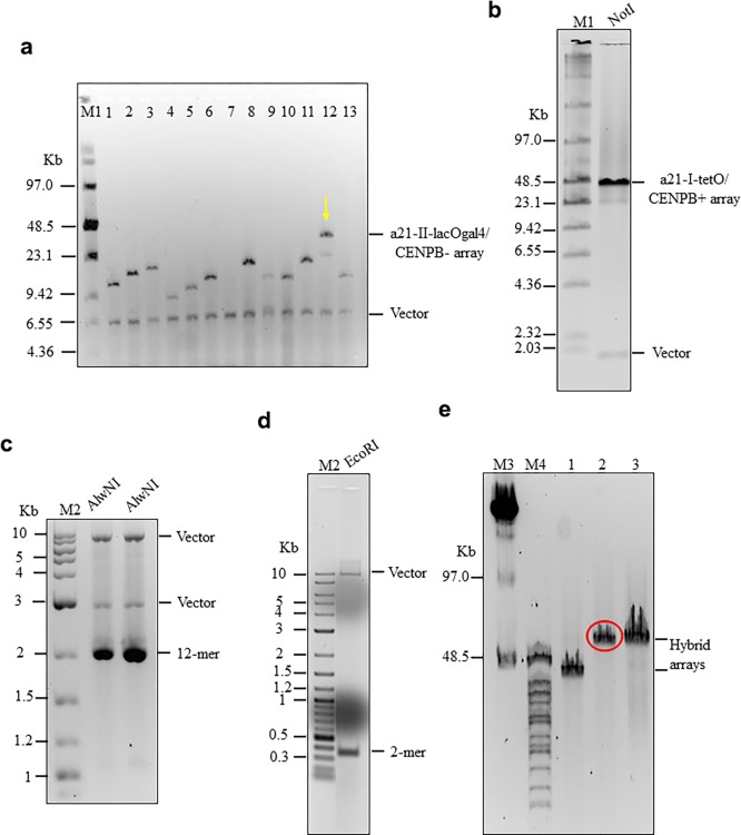
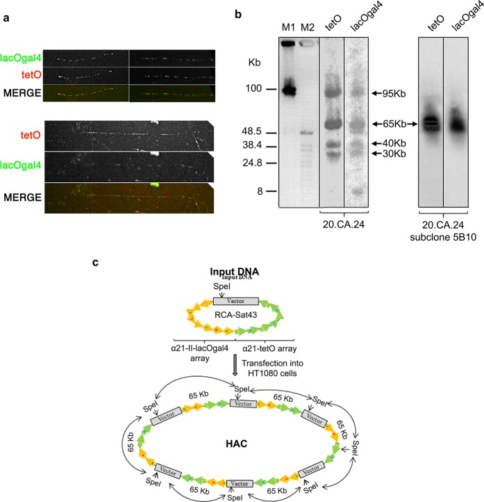
It is generally accepted that chromatin containing the histone H3 variant CENP-A is an epigenetic mark maintaining centromere identity. However, the pathways leading to the formation and maintenance of centromere chromatin remain poorly characterized due to difficulties of analysis of centromeric repeats in native chromosomes. To address this problem, in our previous studies we generated a human artificial chromosome (HAC) whose centromere contains a synthetic alpha-satellite (alphoid) DNA array containing the tetracycline operator, the alphoidtetO-HAC. The presence of tetO sequences allows the specific targeting of the centromeric region in the HAC with different chromatin modifiers fused to the tetracycline repressor. The alphoidtetO-HAC has been extensively used to investigate protein interactions within the kinetochore and to define the epigenetic signature of centromeric chromatin to maintain a functional kinetochore. In this study, we developed a novel synthetic HAC containing two alphoid DNA arrays with different targeting sequences, tetO, lacO and gal4, the alphoidhybrid-HAC. This new HAC can be used for detailed epigenetic engineering studies because its kinetochore can be simultaneously or independently targeted by different chromatin modifiers and other fusion proteins.

Keywords: centromere; chromosome segregation; human artificial chromosome; kinetochore; mitosis.


Conflict of interest statement

The authors declare no competing financial interest.

Figures

Figure 1
Figure 1
Sequence of the alphoid dimer used for construction of the α21-I-tetO array. Both monomers are derived from a chromosome 21 alphoid type I (HOR). One monomer contains a CENP-B box (shaded blue). In the second monomer, the position corresponding to the CENP-B box sequence was replaced by a 42 bp tetO motif (shaded yellow).
Figure 2
Figure 2
Sequence of the alphoid 12-mer used for construction of the α21-II-lacOgal4 array. All monomers were derived from a chromosome 21 alphoid type II array. Four gal4 sequences (two 21 bp in size and two dimers 42 bp in size; shaded green) and four lacO sequences (each 36 bp in size; shaded red) were incorporated into a 12-mer. Fifteen MseI sites are present in the 12-mer.
Figure 3
Figure 3
Schematic representation of construction of synthetic tandem arrays. (a) Step one includes amplification of either a 2,078 bp 21-II-lacOgal4 12-mer or a 343 bp 21-I-tetO dimer by rolling circle amplification (RCA) reaction up to 1–3 kb fragments. (b) Step two includes construction of long alphoid arrays by transformation-associated recombination (TAR) cloning in yeast. The RCA-amplified fragments are cotransformed into yeast cells along with the MluI-linearized RCA-Sat43 vector (the MluI restriction site is located between the hooks). This vector contains a BAC cassette (a BAC replicon and a Clm marker), a YAC cassette (a selectable marker HIS3, a centromere sequence CEN6 from yeast chromosome VI, and yeast origin of replication ARSH4), and a mammalian marker Bsr (the blasticidin gene) that allows the vector to propagate in yeast, bacterial, and mammalian cells and alphoid-specific hooks of 40 bp each (Ebersole et al. 2005). Recombination of the RCA-amplified fragments accompanied by their recombination with the hooks results in the rescue of long arrays as circular YAC/BACs, and 40 kb α21-I-tetO and 40 kb α21-II-lacOgal4 arrays were chosen for further experiments. (c) Construction of the hybrid tetO-CENPB+-lacOgal4-CENPB array. Recombination between the arrays accompanied by their recombination with the vector hooks leads to formation of the hybrid arrays. Ultimately, a molecule containing a 20 kb lacOgal4 array and 25 kb tetO array was chosen for HAC formation.
Figure 4
Figure 4
Hybrid alphoid-DNA array construction. (a) CHEF analysis of 13 BACs with α21-II-lacOgal4 arrays of different size. The BAC DNAs were linearized by AvaII to release a vector part and an array. BAC #12 has an array of ∼40 kb in size. (b) CHEF analysis of the BAC with the α21-I-tetO array of ∼40 kb in size. The BAC DNA was digested by NheI/SpeI to release a vector fragment and the array. (c) Confirmation of the tandem repeat structure of 40 and 60 kb α21-II-lacOgal4 arrays by AlwN1 digestion. CHEF analysis revealed 2,461 bp 12-mer α21-II-lacOgal4 repeat units. (d) Conformation of the tandem repeat structure of a 40 kb α21-I-tetO array by EcoRI digestion. CHEF analysis revealed 343 bp 2-mer 21-I-tetO repeat units. (E) CHEF analysis of the hybrid α21-I-tetO/a21-II-lacOgal4 arrays. (Lane 1) Array consisting of 10 kb of the α21-II-lacOgal4 array and 30 kb of the a21-I-tetO array. (Lane 2) Array consisting of 25 kb α21-II-lacOgal4 array and 30 kb α21-I-tetO array. The array in lane 2 (in red) was chosen for HAC formation. (Lane 3) Array consisting of 15 kb α21-II-lacOgal4 array and 40 kb α21-I-tetO array.
Figure 5
Figure 5
Hybrid HAC formation in HT1080 cells. (a) Representative FISH images of clones containing a HAC (left) and an array integration in an endogenous chromosome (right). (b, c) Screening of blasticidin-resistant clones by FISH. Diagrams represent the frequency of metaphases with HACs (black bars) and array integrations (gray bars) (N = 25) in HT1080 cells without (b) and with (c) CENP-A overexpression. (d) Frequency of HAC-containing clones with (CENP-A OE) and without (CENP-A WT) transient CENP-A overexpression during HAC formation. Only clones with a minimum of 10% metaphases containing HACs were considered as positive (10 vs 33%). (e) Representative two-color oligo-FISH images showing different hybrid HACs (clone 20.CA.07-top and 20.CA.24-bottom) containing tetO (red) and lacOgal4 (green) domains. Images were captured at optimized exposure times to clearly distinguish both signals in either clone (for signal intensity comparison between clones, see Figure S2). (f) Representative image of an HT1080 cell containing HAC clone 20.CA.24 and expressing both lacI-GFP (green) and tetR-mCherry (red) fusion proteins. Merged image (right panel) represents the overlay of GFP, mCherry, and DAPI channels. (g) Frequency of HAC-containing metaphases in the indicated clones containing HACs in the presence of blasticidin and after 30 days after blasticidin washout. The HAC loss rate is indicated in red. (h) Representative immunofluorescence images on metaphase spreads of HAC clone 20.CA.24 and stained with the indicated antibodies. Scale bars = 10 μm.
Figure 6
Figure 6
Structural analysis of the hybrid HAC propagated in human HT1080 cells. (a) Representative fiber-FISH images of clone 20.CA.24 HAC using oligonucleotide probes for tetO (red) and lacO + gal4 sequences (green). Different degrees of fiber stretching are shown (compare upper and lower panels). (b) Genomic DNA possessing the original HAC clone 20.CA.24 (left panel) and its subclone (5B10; right panel) were digested with SpeI endonuclease and separated by CHEF gel electrophoresis (range 10–100 kb). The SpeI recognition site is present once in the RCA-SAT43 vector at position 812 but not in the hybrid array. The transferred membrane was hybridized with radioactively labeled tetO-specific or lacO + gal4-specific probes. The 5B10 subclone has a HAC with a remarkably conserved array. Arrows indicate fragments of 95, 65, 40, and 30 kb in size that are specific to both probes. (c) Diagram illustrating multimerization of input DNA during de novo HAC formation in human HT1080 cells. Input DNA consists of 65 kb hybrid array and 10,209 bp RCA-Sat43 vector sequence.
Figure 7
Figure 7
Epigenetic engineering shows the presence of a two-domain centromere in the alphoidhybrid HAC. (a) ChIP-qPCR analysis of CENP-A levels in HT1080 clone 5B10 containing the alphoidhybrid HAC. The α21-I-tetO (tetO), α21-II-lacOgal4 (lacOgal4) hybridHAC domains, satellite D17Z1 (Chr17), and degenerate satellite type-II (Sat2) repeats were assessed. (b) Representative images of HT1080–5B10 cells expressing the indicated tetR and lacI-fusion proteins and stained with H3K9me3 (second panel) and CENP-A (third panel) antibodies. Merge images represent the overlay of GFP (green), H3K9me3 (blue), and CENP-A (red). (c) Quantification of HAC-associated CENP-A staining in individual cells transfected with the indicated fusion proteins and plotted as A.F.U. Solid bars indicate the medians, and error bars represent the s.e.m. n = two independent experiments for each time point and staining. Asterisks indicate a significant difference (*P < 0.05; **P < 0.01; Mann–Whitney test). Scale bars = 10 μm.
Figure 8
Figure 8
Alphoidhybrid HAC shows epigenetically distinct centromeric domains. (a) Representative images of HT1080–5B10 cells expressing the indicated tetR (first panel) and lacI-fusion proteins (second panel) and stained with antibodies recognizing H3K9me3 (third panel) and CENP-A (fourth panel). Merged images represent the overlay of TMR-SNAP, GFP, and H3K9me3 (MERGE 1; fifth panel) and GFP, H3K9me3 and CENP-A (MERGE 2; sixth panel). (b) Quantification of HAC-associated CENP-A staining in individual cells transfected with the indicated fusion proteins and plotted as A.F.U. Solid bars indicate the medians, and error bars represent the s.e.m. n = two independent experiments for each time point and staining. Asterisks indicate a significant difference (**P < 0.01; Mann–Whitney test). (c) Quantification of alphoidhybrid HAC copy-numbers as determined by counting the GFP and/or TMR-SNAP spot in interphase nuclei of cells transfected with the indicated fusion proteins. Data represent the mean (and s.e.m.) of three independent assays of each time point after doxycycline washout (n = 1,000 nuclei per condition; *P < 0.05, **P < 0.0001; χ2-test). (d) ChIP-qPCR analysis in HT1080–5B10 cells using the indicated antibodies. The α21-I-tetO (tetO), α21-II-lacOgal4 (lacOgal4) hybridHAC domains, the satellite D17Z1 (Chr17), and the degenerate satellite type-II (Sat2) repeats were assessed. Scale bars = 10 μm.

References

    1. Fukagawa T.; Earnshaw W. C. (2014) The centromere: chromatin foundation for the kinetochore machinery,. Dev. Cell 30, 496–508. 10.1016/j.devcel.2014.08.016. - DOI - PMC - PubMed
    1. Schueler M. G.; Sullivan B. A. (2006) Structural and functional dynamics of human centromeric chromatin. Annu. Rev. Genomics Hum. Genet. 7, 301–313. 10.1146/annurev.genom.7.080505.115613. - DOI - PubMed
    1. Earnshaw W. C.; Migeon B. R. (1985) Three related centromere proteins are absent from the inactive centromere of a stable isodicentric chromosome. Chromosoma 92, 290–296. 10.1007/BF00329812. - DOI - PubMed
    1. Sekulic N.; Black B. E. (2012) Molecular underpinnings of centromere identity and maintenance. Trends Biochem. Sci. 37, 220–229. 10.1016/j.tibs.2012.01.003. - DOI - PMC - PubMed
    1. Sullivan B. A.; Schwartz S. (1995) Identification of centromeric antigens in dicentric Robertsonian translocations: CENP-C and CENP-E are necessary components of functional centromeres. Hum. Mol. Genet. 4, 2189–2197. 10.1093/hmg/4.12.2189. - DOI - PubMed

Publication types

MeSH terms