

Optic nerve sheath fenestration: a revised lateral approach for nerve access
- PMID: 29565728
- PMCID: PMC6151165
- DOI: 10.1080/01676830.2018.1452949
Optic nerve sheath fenestration: a revised lateral approach for nerve access
Abstract
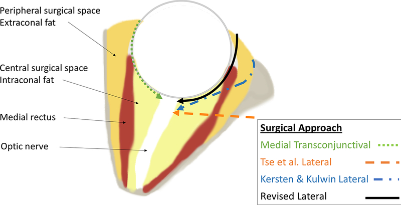
Idiopathic intracranial hypertension (IIH), also known as pseudotumor cerebri, describes a disease of poorly understood pathophysiology with a specific set of signs and symptoms including potentially irreversible and blinding visual loss. Optic nerve sheath fenestration (ONSF) is a well-described surgical treatment for patients with IIH and progressive visual loss despite maximally tolerated medical therapy. A number of optic nerve access procedures have been described including medial transconjunctival, superomedial lid crease, and lateral orbitotomy with and without bone takedown. The purpose of this report is to describe a revised lateral approach for temporal optic nerve access that obviates the need to traverse through the intraconal fat of the central surgical space in the previously described lateral approach techniques.
Keywords: Idiopathic intracranial hypertension; lateral orbitotomy; optic nerve sheath fenestration; pseudotumor cerebri.
Conflict of interest statement
Conflict of Interest:
No conflicting relationship exists for any author.
Figures


Similar articles
-
Superomedial lid crease approach to the medial intraconal space: a new technique for access to the optic nerve and central space.Ophthalmic Plast Reconstr Surg. 2001 Jul;17(4):241-53. doi: 10.1097/00002341-200107000-00003. Ophthalmic Plast Reconstr Surg. 2001. PMID: 11476174
-
Optic nerve sheath fenestration: Current status in France and comparison of 6 different surgical approaches.J Fr Ophtalmol. 2023 Feb;46(2):137-147. doi: 10.1016/j.jfo.2022.07.014. Epub 2022 Dec 21. J Fr Ophtalmol. 2023. PMID: 36564304
-
Optic nerve sheath fenestration in pseudotumor cerebri. A lateral orbitotomy approach.Arch Ophthalmol. 1988 Oct;106(10):1458-62. doi: 10.1001/archopht.1988.01060140622035. Arch Ophthalmol. 1988. PMID: 3052386 Review.
-
Optic Nerve Sheath Fenestration: Current Preferences in Surgical Approach and Biopsy.Ophthalmic Plast Reconstr Surg. 2015 Jul-Aug;31(4):310-2. doi: 10.1097/IOP.0000000000000326. Ophthalmic Plast Reconstr Surg. 2015. PMID: 26168208
-
A 15-year review of secondary and tertiary optic nerve sheath fenestration for idiopathic intracranial hypertension.Orbit. 2018 Aug;37(4):266-272. doi: 10.1080/01676830.2017.1423337. Epub 2018 Jan 9. Orbit. 2018. PMID: 29313398 Review.
Cited by
-
Optic nerve sheath fenestration for visual impairment in cerebral venous diseases.Front Neurol. 2023 Jan 24;14:1065315. doi: 10.3389/fneur.2023.1065315. eCollection 2023. Front Neurol. 2023. PMID: 36761350 Free PMC article.
-
Unilateral Optic Nerve Sheath Fenestration in Idiopathic Intracranial Hypertension: A 6-Month Follow-Up Study on Visual Outcome and Prognostic Markers.Life (Basel). 2021 Jul 31;11(8):778. doi: 10.3390/life11080778. Life (Basel). 2021. PMID: 34440522 Free PMC article.
-
The lateral fornix orbitotomy: a novel surgical corridor to lacrimal gland lesions.Graefes Arch Clin Exp Ophthalmol. 2024 Dec;262(12):3987-3995. doi: 10.1007/s00417-024-06584-w. Epub 2024 Jul 29. Graefes Arch Clin Exp Ophthalmol. 2024. PMID: 39073563
References
-
- Goh KY, Schatz NJ, Glaser JS. Optic nerve sheath fenestration for pseudotumor cerebri. J Neuroophthalmol 1997;17(2):86–91. - PubMed
-
- Mudumbai RC. Optic nerve sheath fenestration: indications, techniques, mechanisms and, results. Int Ophthalmol Clin 2014;54(1):43–9. - PubMed
-
- Radvany MG, Solomon D, Nijjar S, et al. Visual and neurological outcomes following endovascular stenting for pseudotumor cerebri associated with transverse sinus stenosis. J Neuroophthalmol 2013;33(2):117–22. - PubMed
-
- Jiramongkolchai K, Buckley EG, Bhatti MT, et al. Temporary Lumbar Drain as Treatment for Pediatric Fulminant Idiopathic Intracranial Hypertension. J Neuroophthalmol 2017;37(2):126–32. - PubMed
MeSH terms
Grants and funding
LinkOut - more resources
Full Text Sources
Other Literature Sources
Medical