Review
. 2018 Mar 22;9(4):177.
doi: 10.3390/genes9040177.

Role of the Extremolytes Ectoine and Hydroxyectoine as Stress Protectants and Nutrients: Genetics, Phylogenomics, Biochemistry, and Structural Analysis

Affiliations
Review

Role of the Extremolytes Ectoine and Hydroxyectoine as Stress Protectants and Nutrients: Genetics, Phylogenomics, Biochemistry, and Structural Analysis

Laura Czech et al. Genes (Basel). .

Abstract

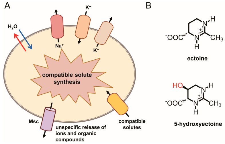
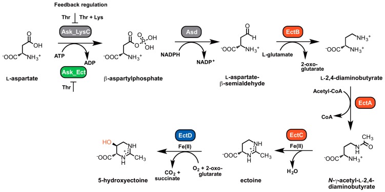
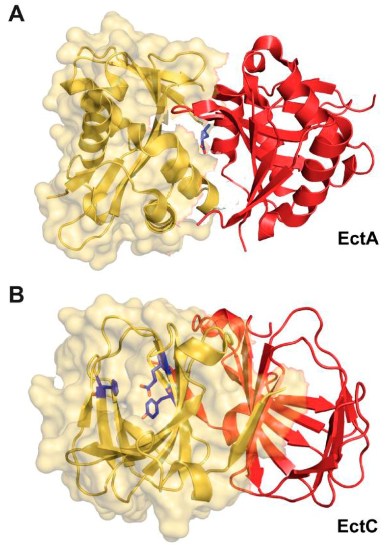
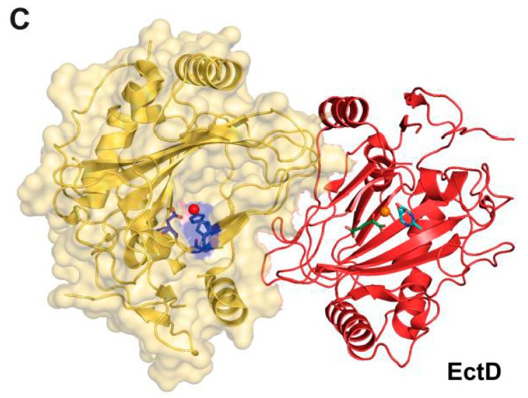
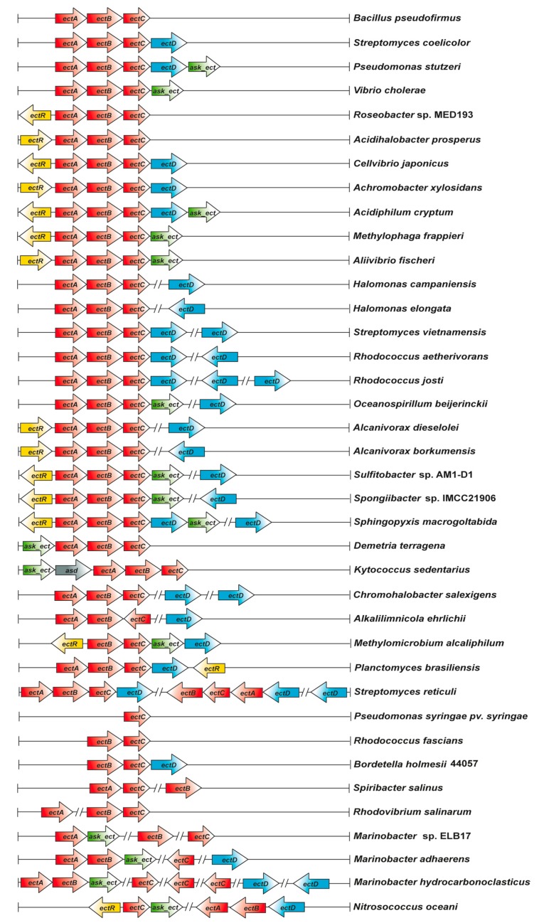
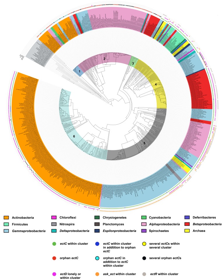
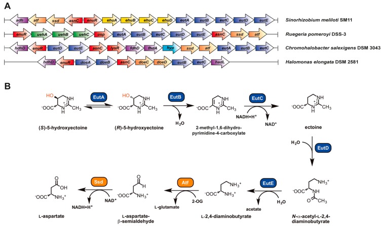
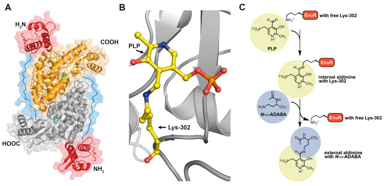
Fluctuations in environmental osmolarity are ubiquitous stress factors in many natural habitats of microorganisms, as they inevitably trigger osmotically instigated fluxes of water across the semi-permeable cytoplasmic membrane. Under hyperosmotic conditions, many microorganisms fend off the detrimental effects of water efflux and the ensuing dehydration of the cytoplasm and drop in turgor through the accumulation of a restricted class of organic osmolytes, the compatible solutes. Ectoine and its derivative 5-hydroxyectoine are prominent members of these compounds and are synthesized widely by members of the Bacteria and a few Archaea and Eukarya in response to high salinity/osmolarity and/or growth temperature extremes. Ectoines have excellent function-preserving properties, attributes that have led to their description as chemical chaperones and fostered the development of an industrial-scale biotechnological production process for their exploitation in biotechnology, skin care, and medicine. We review, here, the current knowledge on the biochemistry of the ectoine/hydroxyectoine biosynthetic enzymes and the available crystal structures of some of them, explore the genetics of the underlying biosynthetic genes and their transcriptional regulation, and present an extensive phylogenomic analysis of the ectoine/hydroxyectoine biosynthetic genes. In addition, we address the biochemistry, phylogenomics, and genetic regulation for the alternative use of ectoines as nutrients.

Keywords: biotechnology; chemical chaperones; crystal structures; enzymes; gene expression; genomics; growth temperature extremes; high salinity; osmotic stress.


Conflict of interest statement

Conflict of Interests: The authors declare no conflict of interest.

Figures

Figure 1
Figure 1
(A) General overview of the microbial salt-out osmostress adaptation strategy. The components, ion fluxes, and compatible solute pools generated via import and synthesis under hyperosmotic conditions [1,2], and the non-specific release of ions and low molecular weight organic compounds via mechanosensitive channels (Msc) under suddenly imposed hypo-osmotic circumstances are depicted [11,20]. (B) Chemical structures of the compatible solutes ectoine and 5-hydroxyectoine.
Figure 2
Figure 2
Routes for ectoine and 5-hydroxyectoine biosynthesis.
Figure 3
Figure 3
Crystal structures of the EctA and EctC ectoine biosynthetic enzymes and that of the ectoine hydroxylase EctD. Dimers of the l-2,4-diaminobutyrate acetyltransferase (EctA), ectoine synthase (EctC), and ectoine hydroxylase (EctD) are depicted. (A) In the crystal structure of the EctA protein from Bordetella parapertussis [Protein Data Bank (PDB) accession code 3D3S] a single molecule of the substrate DABA is bound at the dimer interface. (B) Crystal structure of the EctC protein from Sphingopyxis alaskensis (PDB accession code 5BXX). In one of the dimers, the putative metal-binding residues (Glu57, Tyr85, His93) are highlighted; these protrude into the lumen of the cupin barrel, where the predicted active site of the enzyme is located [176]. (C) Crystal structure of the EctD protein from S. alaskensis (PDB accession code 4Q5O). In the left monomer of the dimer assembly, the three residues (His144, Asp146, His245) coordinating the catalytically important iron (shown as an orange sphere) are highlighted. In the right monomer of the dimer assembly, the position of the co-substrate for the EctD enzyme, 2-oxoglutarate, and the ectoine-derived product 5-hydroxyectoine are depicted relative to that of the ion catalyst [177].
Figure 3
Figure 3
Crystal structures of the EctA and EctC ectoine biosynthetic enzymes and that of the ectoine hydroxylase EctD. Dimers of the l-2,4-diaminobutyrate acetyltransferase (EctA), ectoine synthase (EctC), and ectoine hydroxylase (EctD) are depicted. (A) In the crystal structure of the EctA protein from Bordetella parapertussis [Protein Data Bank (PDB) accession code 3D3S] a single molecule of the substrate DABA is bound at the dimer interface. (B) Crystal structure of the EctC protein from Sphingopyxis alaskensis (PDB accession code 5BXX). In one of the dimers, the putative metal-binding residues (Glu57, Tyr85, His93) are highlighted; these protrude into the lumen of the cupin barrel, where the predicted active site of the enzyme is located [176]. (C) Crystal structure of the EctD protein from S. alaskensis (PDB accession code 4Q5O). In the left monomer of the dimer assembly, the three residues (His144, Asp146, His245) coordinating the catalytically important iron (shown as an orange sphere) are highlighted. In the right monomer of the dimer assembly, the position of the co-substrate for the EctD enzyme, 2-oxoglutarate, and the ectoine-derived product 5-hydroxyectoine are depicted relative to that of the ion catalyst [177].
Figure 4
Figure 4
Diversity of the genetic organization of ectoine and 5-hydroxyectoine biosynthetic gene clusters in microbial genomes.
Figure 5
Figure 5
Phylogenomics of the ectoine synthase. The amino acid sequences of 582 EctC-type proteins were retrieved from microorganisms with fully sequenced genomes, aligned with MAFFT [192] and then used for a clade analysis using the iTOL software [193]. The tree was rooted with a number of microbial cupin-type proteins, a superfamily of proteins [182,183] to which the EctC protein also belongs [176]. The phylogenetic affiliation of the various EctC proteins is depicted in different colors shown in the outer ring, and the color code is explained in the figure. Different groups (1 to 6) in which the EctC-type proteins can be clustered are depicted in the inner colored circle. The dots in the outmost 5 rings depict (from the inside to the outside) if the EctC protein is encoded within an ect biosynthetic gene cluster, if the EctC protein is an orphan, if the pertinent EctC-containing microorganism also possesses the ectoine hydroxylase EctD, if the specialized aspartokinases Ask_Ect is part of the ect cluster, or if the ect gene cluster is affiliated with a gene encoding the EctR regulatory protein.
Figure 6
Figure 6
Genetics and catabolic pathways for the utilization of ectoine and 5-hydroxyectoine as nutrients. (A) Genetic organization of the ectoine/5-hydroxyectoine-catabolic gene cluster in Sinorhizobium meliloti SM11 [265], Ruegeria pomeroyi DSS-3 [263,270], Halomonas elongata DSM 258 [158] and Chromohalobacter salexigens DSM 3043 (predicted from the genome sequence) [274]. In addition to the transporter and catabolic genes discussed in the main text, some of these gene clusters contain genes with yet undefined roles in ectoine catabolism. Their gene products have bioinformatically predicted functions as alcohol dehydrogenase (adh), hydroxyacid dehydrogenase (hdhD), formate dehydrogenases (fdhD, fdhA), haloacid dehalogenase (hadL), transcriptional regulator (lysR) and a hypothetical protein (hyp). (B) Predicted pathway for the catabolism of ectoine and its derivative 5-hydroxyectoine in R. pomeroyi DSS-3. The EutABC-enzymes are predicted to convert 5-hydroxyectoine in a three-step reaction into ectoine. The ectoine ring is subsequently hydrolyzed by the EutD enzyme, resulting in the production of N-α-ADABA, an intermediate, which is then further catabolized to l-aspartate by the EutE, Atf and Ssd enzymes. These data were compiled from the literature [158,263,270]. The ectoine-derived metabolites N-α-ADABA and l-2,4-diaminobutyrate (DABA) serve as inducers for the transcriptional control of the ectoine/5-hydroxyectoine import and catabolic gene clusters by the EnuR regulatory protein [270].
Figure 7
Figure 7
EnuR, a PLP-containing transcriptional regulator of ectoine/5-hydroxyectoine gene clusters. (A) in silico model of the predicted Ruegeria pomeroyi DSS-3 EnuR dimer that was derived from the crystallographic structure of the Bacillus subtilis GabR (PDB accession code 4N0B) [279]. The EnuR model was built with the SWISS-MODEL web server (https://swissmodel.expasy.org/) [286] and visualized using the PyMOL Molecular Graphics System suit (https://pymol.org/2/) [287]. The C-terminal aminotransferase-domains of the EnuR dimer are shown in grey/yellow, the N-terminal DNA-binding domains are represented in red and the flexible linkers connecting these domains are depicted in blue. Each monomer contains a PLP molecule covalently bound via an Schiff base to Lys302 in the aminotransferase domain [263,270]. This internal aldimine [173] is depicted in (B) in a close-up view. (C) Model for the chemistry underlying binding and release of the inducer N-α-ADABA to the PLP-cofactor bound to Lys302 of the EnuR regulator. In the first step, PLP is covalently bound by the side-chain of Lys302 and thus forms an internal aldimine [173]. Upon binding of the inducer N-α-ADABA to PLP, PLP is released from Lys302 and an external aldimine [173] is formed. This sequence of events is envisioned to trigger a conformational change in EnuR, thereby altering its DNA-binding properties. This scheme for inducer binding by EnuR is based upon detailed biochemical and structural analysis of the B. subtilis GabR regulator that uses GABA as its inducer [279,280,281,282,283].

Similar articles

Cited by

References

    1. Kempf B., Bremer E. Uptake and synthesis of compatible solutes as microbial stress responses to high osmolality environments. Arch. Microbiol. 1998;170:319–330. doi: 10.1007/s002030050649. - DOI - PubMed
    1. Bremer E., Krämer R. Coping with osmotic challenges: Osmoregulation through accumulation and release of compatible solutes. In: Storz G., Hengge-Aronis R., editors. Bacterial Stress Responses. ASM Press; Washington, DC, USA: 2000. pp. 79–97.
    1. Wood J.M., Bremer E., Csonka L.N., Krämer R., Poolman B., van der Heide T., Smith L.T. Osmosensing and osmoregulatory compatible solute accumulation by bacteria. Comp. Biochem. Physiol. A Mol. Integr. Physiol. 2001;130:437–460. doi: 10.1016/S1095-6433(01)00442-1. - DOI - PubMed
    1. Csonka L.N. Physiological and genetic responses of bacteria to osmotic stress. Microbiol. Rev. 1989;53:121–147. - PMC - PubMed
    1. Roesser M., Müller V. Osmoadaptation in bacteria and archaea: Common principles and differences. Environ. Microbiol. 2001;3:743–754. doi: 10.1046/j.1462-2920.2001.00252.x. - DOI - PubMed

LinkOut - more resources