. 2018 Mar 22;13(3):e0194868.
doi: 10.1371/journal.pone.0194868. eCollection 2018.

MERS-CoV pathogenesis and antiviral efficacy of licensed drugs in human monocyte-derived antigen-presenting cells

Affiliations

MERS-CoV pathogenesis and antiviral efficacy of licensed drugs in human monocyte-derived antigen-presenting cells

Yu Cong et al. PLoS One. .

Abstract

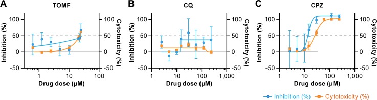
Middle East respiratory syndrome coronavirus (MERS-CoV) presents an emerging threat to public health worldwide by causing severe respiratory disease in humans with high virulence and case fatality rate (about 35%) since 2012. Little is known about the pathogenesis and innate antiviral response in primary human monocyte-derived macrophages (MDMs) and dendritic cells (MDDCs) upon MERS-CoV infection. In this study, we assessed MERS-CoV replication as well as induction of inflammatory cytokines and chemokines in MDMs and immature and mature MDDCs. Immature MDDCs and MDMs were permissive for MERS-CoV infection, while mature MDDCs were not, with stimulation of proinflammatory cytokine and chemokine upregulation in MDMs, but not in MDDCs. To further evaluate the antiviral activity of well-defined drugs in primary antigen presenting cells (APCs), three compounds (chloroquine, chlorpromazine and toremifine), each with broad-spectrum antiviral activity in immortalized cell lines, were evaluated in MDMs and MDDCs to determine their antiviral effect on MERS-CoV infection. While chloroquine was not active in these primary cells, chlorpromazine showed strong anti-MERS-CoV activity, but it was associated with high cytotoxicity narrowing the potential window for drug utilization. Unlike in established cells, toremifene had marginal activity when tested in antigen presenting cells, with high apparent cytotoxicity, also limiting its potential as a therapeutic option. These results demonstrate the value of testing drugs in primary cells, in addition to established cell lines, before initiating preclinical or clinical studies for MERS treatment and the importance of carefully assessing cytotoxicity in drug screen assays. Furthermore, these studies also highlight the role of APCs in stimulating a robust protective immune response to MERS-CoV infection.


Conflict of interest statement

Competing Interests: The authors have declared that they have no competing interests.

Figures

Fig 1
Fig 1. Differentiation of macrophages and dendritic cells from peripheral blood mononuclear cells.
CD14+ monocytes were isolated from human peripheral blood mononuclear cells (PBMCs). Cells were then differentiated to monocyte-derived macrophages (MDMs) by adding macrophage-colony stimulating factor (M-CSF) or to immature monocyte-derived dendritic cells (MDDCs) by adding granulocyte macrophage-colony stimulating factor (GM-CSF) and interleukin-4 (IL-4). Immature MDDCs were exposed to MDDC maturation cocktail (R-10 medium containing 10 ng/ml of tumor necrosis factor (TNF)-α, 10 ng/ml of IL-1β, 15 ng/ml of IL-6, 1 μg/ml of prostaglandin E2 (PEG2), 20 ng/ml of GM-CSF, 10 ng/ml of IL-4) overnight for maturation. MDMs and MDDCs were characterized by flow cytometry for expression of major cell-surface markers. Using these protocols, the MDMs are predominantly CD14+, CD11b+, HLA-DR+, CD169+, CD163+, and CD86+. The MDDCs are predominantly CD14-, CD11c+, HLA-DR+, CD80+, and CD86+. Mature MDDCs are CD83+, while immature MDDCs are CD83-.
Fig 2
Fig 2. Viral titers from supernatants of MERS-CoV-infected macrophages and dendritic cells via plaque assay.
(A) Macrophages (MDMs), (B) immature monocyte-derived dendritic cells (MDDCs), and (C) mature MDDCs were infected at an MOI = 2 with MERS-CoV Jordan variant. Culture supernatants were collected at indicated times post-inoculation to detect viral titers. Each line represents cells from each individual donor. Titrations were performed in triplicate with data points representing the mean of the triplicate values.
Fig 3
Fig 3. Cytokine and chemokine responses to MERS-CoV infection from certain innate immune primary cells.
(A) Human macrophages (MDMs) (B), immature monocyte-derived (MDDCs), and (C) mature MDDCs were inoculated with MERS-CoV Jordan variant at an MOI = 2 or subjected to mock infection. From cell culture supernatants, the fold change in immunoregulatory cytokines and chemokines relative to that of mock-infected cells incubated for the same period of time, were determined by multiplex assay. All data points represent triplicate biological replicates from four donors.
Fig 4
Fig 4. Antiviral activity of three compounds on MERS-CoV infection in MDMs.
(A) Effect of pretreatment of monocyte-derived macrophages (MDMs) with toremifene (TOMF), (B) chloroquine (CQ), and (C) chlorpromazine (CPZ) prior to MERS-CoV exposure on subsequent infection. The effect of these compounds on the cytotoxicity of macrophages without virus was also studied. MERS-CoV S protein was detected in infected cells using an immunofluorescence assay. The antigen was detected with a rabbit polyclonal antibody to S protein followed by staining with Alexa Fluor 594-conjugated goat anti-rabbit antibody. Percent inhibition of MERS-CoV infection is shown in blue, and percent cytotoxicity of the compounds without virus is shown in orange. Dotted gray line indicates half maximal effective concentrations (EC50) and 50% cytotoxicity concentrations (CC50). The selectivity index (SI) is defined as CC50÷EC50. Results are representative of 2 individual experiments with 3 replicates in each experiment (means ± standard error of the means [SEM]).
Fig 5
Fig 5. Antiviral activity of three compounds on MERS-CoV infection in MDMs and immature MDDCs.
(A) Monocyte-derived macrophages (MDMs), (B) immature dendritic cells (MDDCs) and (C) Vero E6 cells were pretreated with toremifene (TOMF), chloroquine (CQ), and chlorpromazine (CPZ) for 1 h followed by inoculation with MERS-CoV Jordan variant at an MOI = 0.1. Supernatants collected at 72 h post infection (hpi) for APCs and 48 hpi for Vero E6 cells were inoculated onto Vero E6 cells pre-seeded overnight in 96 well plates and incubated for 6 days. Wells with cytopathic effects were counted, and TCID50 was determined using the Reed-Muench method. Cytotoxicity was measured in parallel using luminescent cell viability assay at 48 h after compounds addition to the mock-infected MDMs and immature MDDCs. Antiviral activity is shown in gray and cytotoxicity is shown in orange. Results are representative of 2 individual experiments with 3 replicates in each (means ± standard error of the means [SEM]).

References

    1. Zaki AM, van Boheemen S, Bestebroer TM, Osterhaus AD, Fouchier RA. Isolation of a novel coronavirus from a man with pneumonia in Saudi Arabia. N Engl J Med. 2012;367(19):1814–20. Epub 2012/10/19. doi: 10.1056/NEJMoa1211721 . - DOI - PubMed
    1. de Groot RJ, Baker SC, Baric RS, Brown CS, Drosten C, Enjuanes L, et al. Middle East respiratory syndrome coronavirus (MERS-CoV): announcement of the Coronavirus Study Group. Journal of virology. 2013;87(14):7790–2. Epub 2013/05/17. doi: 10.1128/JVI.01244-13 ; PubMed Central PMCID: PMCPmc3700179. - DOI - PMC - PubMed
    1. World Health Organization. Middle East respiratory syndrome coronavirus (MERS-CoV)—Saudi Arabia Disease outbreak news. Geneva, CH: World Health Organization; 2017. [cited 2017 11 September]. Available from: http://www.who.int/csr/don/6-september-2017-mers-saudi-arabia/en/.
    1. Almario CV, Chey W, Kaung A, Whitman C, Fuller G, Reid M, et al. Computer-generated vs. physician-documented history of present illness (HPI): results of a blinded comparison. Am J Gastroenterol. 2015;110(1):170–9. doi: 10.1038/ajg.2014.356 ; PubMed Central PMCID: PMC4289091. - DOI - PMC - PubMed
    1. Haagmans BL, Al Dhahiry SH, Reusken CB, Raj VS, Galiano M, Myers R, et al. Middle East respiratory syndrome coronavirus in dromedary camels: an outbreak investigation. Lancet Infect Dis. 2014;14(2):140–5. doi: 10.1016/S1473-3099(13)70690-X . - DOI - PMC - PubMed

Publication types

Substances