

Transcranial magnetic motor evoked potentials and magnetic resonance imaging findings in paraplegic dogs with recovery of motor function
- PMID: 29566440
- PMCID: PMC5980462
- DOI: 10.1111/jvim.15058
Transcranial magnetic motor evoked potentials and magnetic resonance imaging findings in paraplegic dogs with recovery of motor function
Abstract
Background: Transcranial magnetic motor evoked potentials (TMMEP) are associated with severity of clinical signs and magnetic resonance imaging (MRI) findings in dogs with spinal cord disease.
Hypothesis: That in initially paraplegic dogs with thoracolumbar intervertebral disc herniation (IVDH), MRI findings before surgery and TMMEPs obtained after decompressive surgery are associated with long-term neurological status and correlate with each other.
Animals: Seventeen client-owned paraplegic dogs with acute thoracolumbar IVDH.
Methods: Prospective observational study. TMMEPs were obtained from pelvic limbs and MRI (3T) of the spinal cord was performed at initial clinical presentation. Follow-up studies were performed ≤ 2 days after reappearance of motor function and 3 months later. Ratios of compression length, intramedullary hyperintensities' length (T2-weighted hyperintensity length ratio [T2WLR]), and lesion extension (T2-weighted-lesion extension ratio) in relation to the length of the 2nd lumbar vertebral body were calculated.
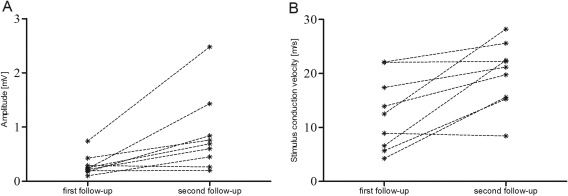
Results: TMMEPs could be elicited in 10/17 (59%) dogs at 1st and in 16/17 (94%) dogs at 2nd follow-up. Comparison of TMMEPs of 1st and 2nd follow-up showed significantly increased amplitudes (median from 0.19 to 0.45 mV) and decreased latencies (from 69.38 to 40.26 ms; P = .01 and .001, respectively). At 2nd follow-up latencies were significantly associated with ambulatory status (P = .024). T2WLR obtained before surgery correlated with latencies at 2nd follow-up (P = .04).
Conclusions: TMMEP reflect motor function recovery after severe spinal cord injury.
Keywords: canine; magnetic resonance imaging; spinal cord injury; therapy monitoring; transcranial magnetic stimulation.
Copyright © 2018 The Authors. Journal of Veterinary Internal Medicine published by Wiley Periodicals, Inc. on behalf of the American College of Veterinary Internal Medicine.
Figures


References
-
- Duval J, Dewey C, Roberts R, Aron D. Spinal cord swelling as a myelographic indicator of prognosis: A retrospective study in dogs with intervertebral disc disease and loss of deep pain perception. Vet Surg. 1996;25:6–12. - PubMed
-
- Jeffery ND, Blakemore WF. Spinal cord injury in small animals. 1. Mechanisms of spontaneous recovery. Vet Rec. 1999;144:407–413. - PubMed
-
- Ferreira AJA, Correia JHD, Jaggy A. Thoracolumbar disc disease in 71 paraplegic dogs: influence of rate of onset and duration of clinical signs on treatment results. J Small Anim Pract. 2002;43:158–163. - PubMed
-
- Olby N, Levine J, Harris T, et al. Long‐term functional outcome of dogs with severe injuries of the thoracolumbar spinal cord: 87 cases (1996–2001). J Am Vet Med Assoc. 2003;222:762–769. - PubMed
-
- Mayhew PD, McLear RC, Ziemer LS, et al. Risk factors for recurrence of clinical signs associated with thoracolumbar intervertebral disk herniation in dogs: 229 cases (1994–2000). J Am Vet Med Assoc. 2004;225:1231–1236. - PubMed
MeSH terms
LinkOut - more resources
Full Text Sources
Other Literature Sources
