

The in vitro modulation of steroidogenesis by inflammatory cytokines and insulin in TM3 Leydig cells
- PMID: 29566712
- PMCID: PMC5863825
- DOI: 10.1186/s12958-018-0341-2
The in vitro modulation of steroidogenesis by inflammatory cytokines and insulin in TM3 Leydig cells
Abstract
Background: Cytokines and hormones, including insulin, are known to modulate the hypothalamic-pituitary-testes axis and steroidogenesis, both centrally and peripherally. In the context of chronic inflammation and hyperinsulinaemia mediating male hypogonadism associated with obesity, metabolic syndrome and type 2 diabetes mellitus, these mechanisms are poorly understood and the impact of cytokines and insulin on Leydig cell steroidogenesis has not been fully elicited. This study aimed to further investigate the in vitro impact of TNFα, IL1ß, IL6, IL8 and insulin on Leydig cell function and steroidogenesis.
Methods: hCG-stimulated TM3 Leydig cells were exposed to various concentrations of TNFα, IL1ß, IL6, IL8 (100 ng/ml, 10 ng/ml, 1 ng/ml and 0.1 ng/ml) and insulin (10 ng/ml, 1 ng/ml, 0.1 ng/ml and 0.01 ng/ml) in optimal cell culture conditions over 48 h. Cell viability (XTT) and testosterone and progesterone concentrations (ELISA) were assessed using standardised laboratory techniques.
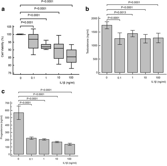
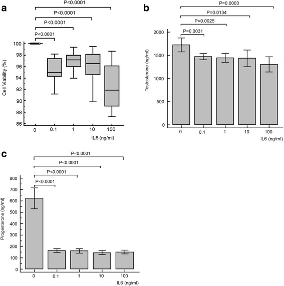
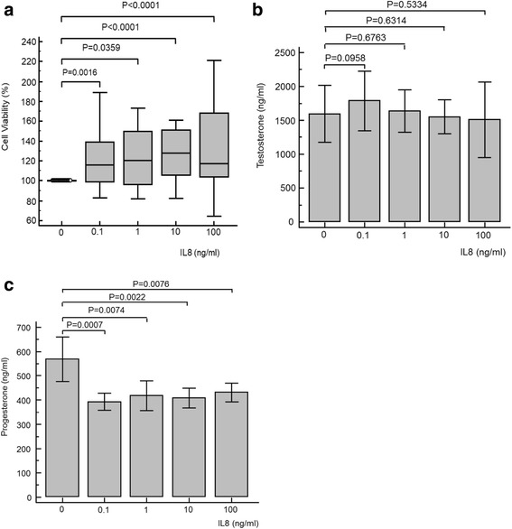
Results: TNFα significantly decreased cell viability and progesterone and testosterone concentrations in a dose-dependent relationship. IL1ß and IL6 had a subtle but significant negative effect on cell viability and testosterone concentrations, with a marked significant decrease in progesterone concentration at all concentrations investigated. IL8 showed an increase in cell viability, with no significant effect on testosterone concentrations alongside a significant decrease in progesterone concentrations. Insulin significantly increased cell viability and testosterone concentrations in a dose dependent relationship, but interestingly significantly decreased progesterone concentrations.
Conclusions: The inflammatory cytokines TNFα, IL1β and IL6 cause a dose dependent decline in steroidogenesis in TM3 Leydig cells. These results suggest that chronic inflammation may downregulate steroidogenesis in males via direct modulation of Leydig cell function. However, IL8 may stimulate TM3 Leydig cell growth. Insulin is associated with a dose-dependent increase in testosterone synthesis, with a significant decline in progesterone synthesis. With the phenomenon of insulin resistance, the literature is unclear on the potential role of hyperinsulinaemia in steroidogenesis. Further studies are warranted in order to fully elicit the molecular mechanisms and interactions of these molecules on male steroidogenesis.
Keywords: Cytokines; IL1β; IL6; IL8; Insulin; Leydig cells; Progesterone; Steroidogenesis; TNFα; Testosterone.
Conflict of interest statement
Ethics approval and consent to participate
Not applicable.
Consent for publication
Not applicable.
Competing interests
The authors declare that they have no competing interests in the study.
Publisher’s Note
Springer Nature remains neutral with regard to jurisdictional claims in published maps and institutional affiliations.
Figures


Similar articles
-
Prolactin and MA-10 Leydig cell steroidogenesis: biphasic effects of prolactin and signal transduction.Endocrinology. 1996 Dec;137(12):5509-18. doi: 10.1210/endo.137.12.8940378. Endocrinology. 1996. PMID: 8940378
-
The Effect of Aqueous Lessertia frutescens Extract on TM3 Leydig Cells Exposed to TNF-α in vitro.Front Biosci (Landmark Ed). 2023 Sep 24;28(9):213. doi: 10.31083/j.fbl2809213. Front Biosci (Landmark Ed). 2023. PMID: 37796713
-
Lead affects steroidogenesis in rat Leydig cells in vivo and in vitro.Toxicology. 1995 Nov 20;103(1):53-62. doi: 10.1016/0300-483x(95)03107-q. Toxicology. 1995. PMID: 8525490
-
Expression of functional leptin receptors in rodent Leydig cells.Endocrinology. 1999 Nov;140(11):4939-47. doi: 10.1210/endo.140.11.7088. Endocrinology. 1999. PMID: 10537117
-
Immune Cells as Critical Regulators of Steroidogenesis in the Testis and Beyond.Front Endocrinol (Lausanne). 2022 Apr 28;13:894437. doi: 10.3389/fendo.2022.894437. eCollection 2022. Front Endocrinol (Lausanne). 2022. PMID: 35573990 Free PMC article. Review.
Cited by
-
A Review on the Impact of Oxidative Stress and Medicinal Plants on Leydig Cells.Antioxidants (Basel). 2023 Aug 4;12(8):1559. doi: 10.3390/antiox12081559. Antioxidants (Basel). 2023. PMID: 37627554 Free PMC article. Review.
-
Downregulating testosterone levels enhance immunotherapy efficiency.Oncoimmunology. 2021 Sep 27;10(1):1981570. doi: 10.1080/2162402X.2021.1981570. eCollection 2021. Oncoimmunology. 2021. PMID: 34595060 Free PMC article.
-
Protective effects of ginseng stem-leaf saponins on D-galactose-induced reproductive injury in male mice.Aging (Albany NY). 2021 Mar 10;13(6):8916-8928. doi: 10.18632/aging.202709. Epub 2021 Mar 10. Aging (Albany NY). 2021. PMID: 33714944 Free PMC article.
-
Characterization of the structural, oxidative, and immunological features of testis tissue from Zucker diabetic fatty rats.Open Life Sci. 2022 Nov 7;17(1):1383-1397. doi: 10.1515/biol-2022-0495. eCollection 2022. Open Life Sci. 2022. PMID: 36405233 Free PMC article.
-
Protective effects of Anthocleista djalonensis A. Chev root extracts against induced testicular inflammation and impaired spermatogenesis in adult rats.Mol Biol Rep. 2019 Dec;46(6):5983-5994. doi: 10.1007/s11033-019-05033-w. Epub 2019 Aug 19. Mol Biol Rep. 2019. PMID: 31428909
References
-
- Pasquali R, Casimirri F, De Iasio R, Mesini P, Boschi S, Chierici R, Flamia R, Biscotti M, Vicennati V. Insulin regulates testosterone and sex hormone-binding globulin concentrations in adult normal weight and obese men. J Clin Endocrinol Metab. 1995;80:654–658. - PubMed
MeSH terms
Substances
LinkOut - more resources
Full Text Sources
Other Literature Sources
Medical
