. 2018 Mar 21;97(6):1284-1298.e7.
doi: 10.1016/j.neuron.2018.02.015.

Tau Kinetics in Neurons and the Human Central Nervous System

Affiliations

Tau Kinetics in Neurons and the Human Central Nervous System

Chihiro Sato et al. Neuron. .

Erratum in

  • Tau Kinetics in Neurons and the Human Central Nervous System.
    Sato C, Barthélemy NR, Mawuenyega KG, Patterson BW, Gordon BA, Jockel-Balsarotti J, Sullivan M, Crisp MJ, Kasten T, Kirmess KM, Kanaan NM, Yarasheski KE, Baker-Nigh A, Benzinger TLS, Miller TM, Karch CM, Bateman RJ. Sato C, et al. Neuron. 2018 May 16;98(4):861-864. doi: 10.1016/j.neuron.2018.04.035. Neuron. 2018. PMID: 29772204 Free PMC article. No abstract available.

Abstract

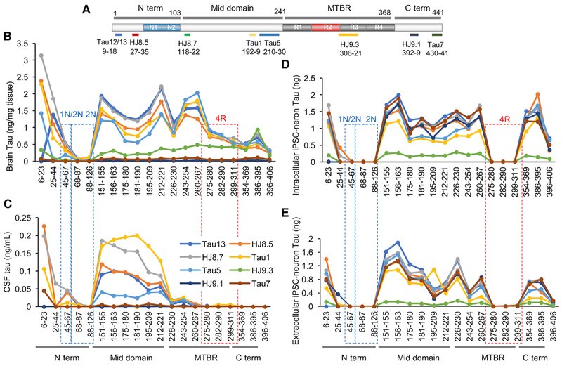
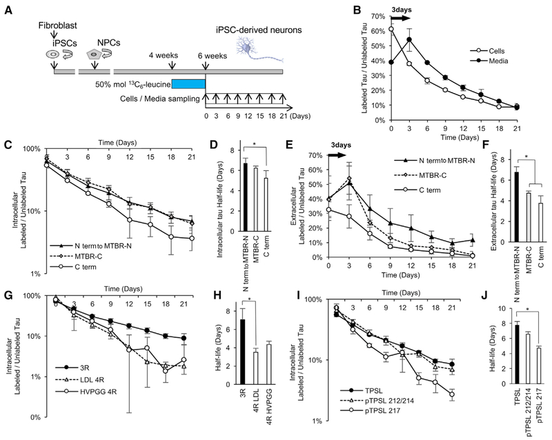
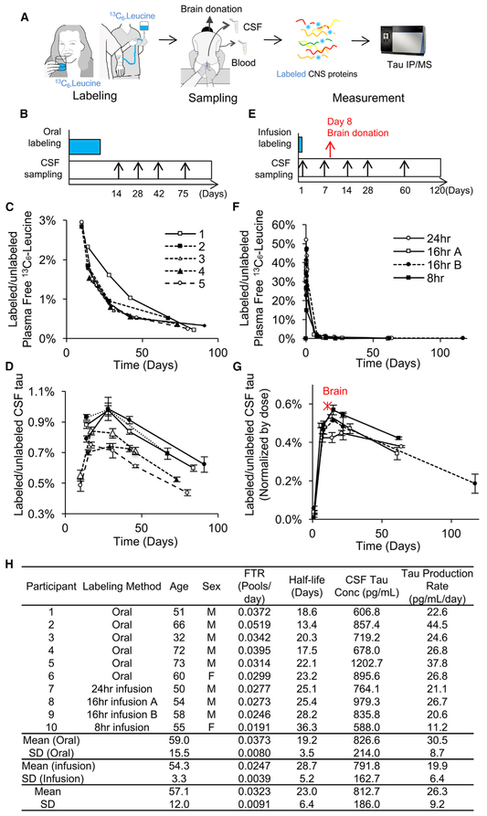
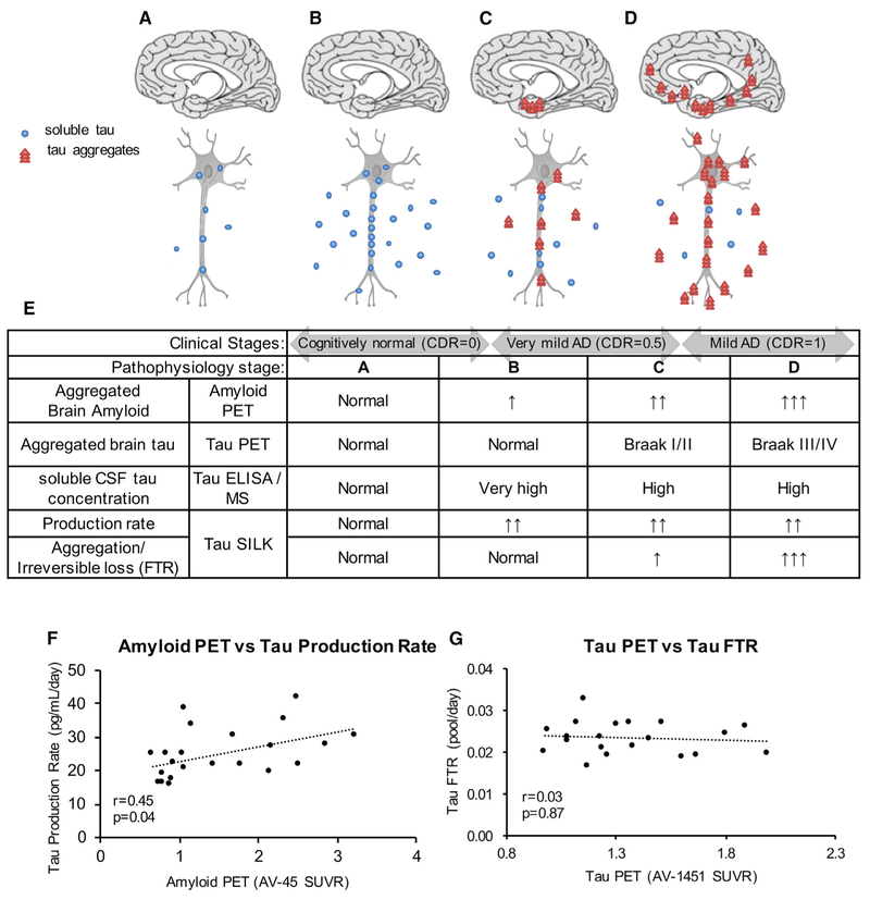
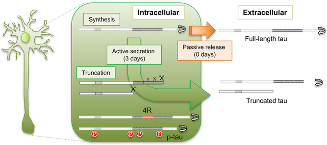
We developed stable isotope labeling and mass spectrometry approaches to measure the kinetics of multiple isoforms and fragments of tau in the human central nervous system (CNS) and in human induced pluripotent stem cell (iPSC)-derived neurons. Newly synthesized tau is truncated and released from human neurons in 3 days. Although most tau proteins have similar turnover, 4R tau isoforms and phosphorylated forms of tau exhibit faster turnover rates, suggesting unique processing of these forms that may have independent biological activities. The half-life of tau in control human iPSC-derived neurons is 6.74 ± 0.45 days and in human CNS is 23 ± 6.4 days. In cognitively normal and Alzheimer's disease participants, the production rate of tau positively correlates with the amount of amyloid plaques, indicating a biological link between amyloid plaques and tau physiology.

Keywords: Alzheimer’s disease; PET; SILK; amyloid; human; induced pluripotent stem cell; isoform; phosphorylation; positron emission tomography; production rate; stable isotope labeling kinetics; tau.


Conflict of interest statement

DECLARATION OF INTERESTS

Washington University and R.J.B. have equity ownership interest in C2N Diagnostics and may receive royalty income based on technology licensed by Washington University to C2N Diagnostics. R.J.B. receives income from C2N Diagnostics for serving on the Scientific Advisory Board, and B.W.P. receives consultation income from C2N Diagnostics. T.L.S.B. participates in research sponsored by Avid Radiopharmaceuticals, Eli Lilly, Roche, Johnson & Johnson, and Biogen. Washington University has submitted the U.S. non-provisional patent application “Methods for measuring the metabolism of CNS derived biomolecules in vivo,” serial #12/267,974 (R.J.B. and K.G.M.), and “Tau kinetics measurements,” serial #CA 2962969 (R.J.B., T.M.M., K.G.M., and C.S.). The remaining co-authors have no conflicts to disclose.

Figures

Figure 1.
Figure 1.. Tau Profiles in the Human CNS and Neurons
(A)Schematic of the longest tau isoform (2N4R), fragments detected by MS, and epitopes of tau antibodies. (B–E) Quantitation of tau peptides in the human brain (B), normal control CSF (C), cell lysates (D), and media (E) from iPSC-derived neurons. Brain tau (B) and cellular tau (D) are a mixture of full-length and C-terminally truncated tau. CSF tau (C) is truncated at the end of the mid-domain (between amino acids 222 and 225). Extracellular tau from iPSC-derived neurons (E) is primarily C-terminally truncated. Intracellular tau from iPSC-derived neurons was diluted to match extracellular tau level. Red dashed lines indicate 4R-containing peptides in the microtubule-binding region (MTBR). Blue dashed lines indicate 1N- and 2N-containing peptides. Data are represented as mean ± SD. See also Figures S1–S3.
Figure 2.
Figure 2.. Differential Tau Kinetics of Tau Isoforms in iPSC-Derived Neurons
(A) A diagram illustrating the in vitro tau SILK protocol. iPSCs were converted into neural precursor cells (NPCs) and differentiated into iPSC-derived neurons for 4 weeks. iPSC-derived neurons were pre-labeled with 50 mol % (100% TTR) 13C6-leucine for 2 weeks from week 4 to 6 (pulse). iPSC-derived neurons were cultured in label-free media for 21 days (chase). Cells and media were collected every 3 days. (B) Kinetic profiles of tau (TPSLPTPPTR) peptides in the cells and in the media. There is a 3-day delay in the newly translated tau released from the intracellular to extracellular space. (C) Kinetic profiles of intracellular peptides. (D) C terminus (C term) peptides have faster turnover and significantly shorter half-lives (p = 0.0003; F = 9.48; Df = 39). (E) Kinetic profiles of extracellular peptides. N terminus, mid-domain, and N terminus of microtubule-binding region (N term to MTBR-N) peptides have similar kinetic profiles. Full-length tau containing C terminus (C term) does not have a 3-day time delay and has a similar kinetic profile as intracellular full-length tau. C terminus of microtubule-binding region (MTBR-C) is released with 3-day time delay and has a shorter half-life than N term to MTBR-N tau (p < 0.0001; F = 18.28; Df = 30). (F) MTBR-C and C term tau peptides have significantly shorter half-lives than N term to MTBR-N tau (p = 0.0033; F = 18.28, Df = 30). (G and H) 4R tau isoform-specific peptides (LDL and HVPGG; see Table 1 for abbreviations) have faster turnover and shorter half-lives in the cell lysates compared to the 3R (p = 0.0007; F = 5.455; Df = 29). (I and J) Phosphorylated tau at T217 (pTPSL 217) has a faster turnover and shorter half-life compared to unphosphorylated tau in the cell lysate (p = 0.0057; F = 8.5; Df = 10). Significance was determined via one-way ANOVA with Tukey’s post hoc test. *p % 0.05. Data are represented as mean ± SD. See also Tables 1 and 2 and Figures S4 and S5.
Figure 3.
Figure 3.. Tau Kinetics in the Human CNS
(A) A diagram illustrating the tau SILK method in humans. (B–G) Six participants were orally labeled for 10 days (B–D), and four participants were labeled with 24, 16 (A and B are two different participants), or 8 hr i.v. infusion (E–G) with 13C6-leucine. Examples of a sampling timeline for oral labeling (B) and infusion labeling (E) protocols. CSF samples were assessed using tau SILK analyses and free leucine in the plasma was measured by gas chromatography-MS (GC-MS) (C and F). 13C6 leucine-labeled tau in CSF was measured in triplicate using LC-MS (D and G) and was normalized by the given dose to match 16 hr infusion in (G). TTR of the human brain 8 days after 800 mg i.v. bolus labeling is plotted as a red star in (G), indicating similar labeling kinetics of brain and CSF in humans. (H) Age, sex, FTR, half-life of CNS tau, CSF tau concentration measured by MS, tau production rate, and CV% of MS triplicate measurements. Data are represented as mean ± SD. See also Figure S6.
Figure 4.
Figure 4.. Tau Kinetics in AD
(A–E) Proposed hypothesis of tau turnover in AD pathogenesis. Schematics (A–D) of tau production and aggregation as disease progresses and a table summary (E) of the changes in tau measured by tau PET imaging, tau ELISA/MS, and tau SILK. Stage A: physiological tau secretion. Defined to be normal. Stage B: increased tau production and soluble tau secretion, initiated by amyloid beta toxicity or other causes. Stage C: increased aggregated tau and decreasing elevated soluble CSF tau. Stage D: trans-synaptic spreading of aggregated tau. (F and G) Tau production rate positively correlates with amyloid PET measure by AV-45 (F). Tau FTR does not correlate with tau PET (G). Spearman correlation (r and p value) was calculated to determine each association. See also Table 3 and Figures S7 and S8.
Figure 5.
Figure 5.. Tau Truncation and Release Are Regulated
Our findings support an active process of translation, processing, and release of tau that occurs over 3 days, and specific increase in turnover of modified tau associated with tauopathy-based diseases such as AD, PSP, and other tauopathies. Diagram showing proposed hypotheses for truncation and secretion of tau. Full-length tau is passively released (orange arrow, no delay). Truncated tau is actively released to the extracellular space (green arrow, 3-day delay). Tau is truncated at multiple sites, including between MTBR-C (Figure 2) and C terminus, and at the end of mid-domain. We propose that longer, truncated forms including MTBR-C are more readily secreted but are more rapidly turned over (gray protein conversion). Within the cell, 4R isoforms and phosphorylated tau undergo more rapid intracellular turnover (gray proteins with red modifications).

Comment in

References

    1. Baker M, Litvan I, Houlden H, Adamson J, Dickson D, Perez-Tur J, Hardy J, Lynch T, Bigio E, and Hutton M (1999). Association of an extended haplotype in the tau gene with progressive supranuclear palsy. Hum. Mol. Genet 8, 711–715. - PubMed
    1. Ban H, Nishishita N, Fusaki N, Tabata T, Saeki K, Shikamura M, Takada N, Inoue M, Hasegawa M, Kawamata S, and Nishikawa S (2011). Efficient generation of transgene-free human induced pluripotent stem cells (iPSCs) by temperature-sensitive Sendai virus vectors. Proc. Natl. Acad. Sci. USA 108, 14234–14239. - PMC - PubMed
    1. Barthélemy NR, Fenaille F, Hirtz C, Sergeant N, Schraen-Maschke S, Vialaret J, Buée L, Gabelle A, Junot C, Lehmann S, and Becher F (2016). Tau protein quantification in human cerebrospinal fluid by targeted mass spectrometry at high sequence coverage provides insights into its primary structure heterogeneity. J. Proteome Res 15, 667–676. - PubMed
    1. Barthélemy NR, Bateman RJ, Marin P, Becher F, Sato C, Lehmann S, and Gabelle A (2017). Tau hyperphosphorylation on T217 in cerebrospinal fluid is specifically associated to amyloid-β pathology. bioRxiv. 10.1101/226977. - DOI
    1. Bateman RJ, Munsell LY, Morris JC, Swarm R, Yarasheski KE, and Holtzman DM (2006). Human amyloid-beta synthesis and clearance rates as measured in cerebrospinal fluid in vivo. Nat. Med 12, 856–861. - PMC - PubMed

Publication types

MeSH terms