. 2018 Jun:167:121-131.
doi: 10.1016/j.biomaterials.2018.03.003. Epub 2018 Mar 13.

Inducing hair follicle neogenesis with secreted proteins enriched in embryonic skin

Affiliations

Inducing hair follicle neogenesis with secreted proteins enriched in embryonic skin

Sabrina Mai-Yi Fan et al. Biomaterials. 2018 Jun.

Abstract

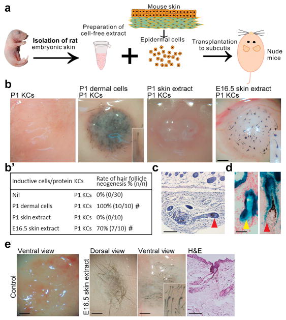
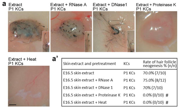
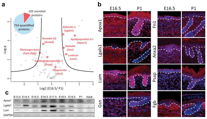
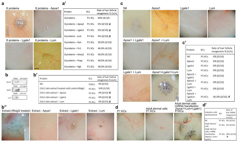
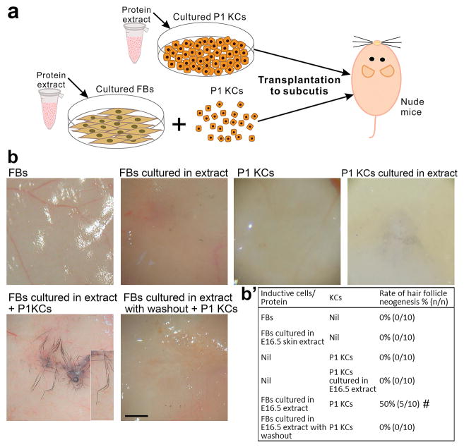
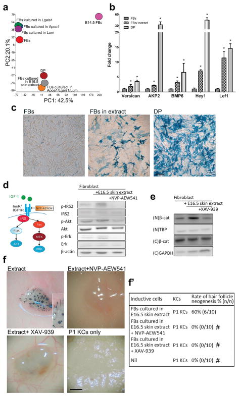
Organ development is a sophisticated process of self-organization. However, despite growing understanding of the developmental mechanisms, little is known about how to reactivate them postnatally for regeneration. We found that treatment of adult non-hair fibroblasts with cell-free extract from embryonic skin conferred upon them the competency to regenerate hair follicles. Proteomics analysis identified three secreted proteins enriched in the embryonic skin, apolipoprotein-A1, galectin-1 and lumican that together were essential and sufficient to induce new hair follicles. These 3 proteins show a stage-specific co-enrichment in the perifolliculogenetic embryonic dermis. Mechanistically, exposure to embryonic skin extract or to the combination of the 3 proteins altered the gene expression to an inductive hair follicle dermal papilla fibroblast-like profile and activated Igf and Wnt signaling, which are crucial for the regeneration process. Therefore, a cocktail of organ-specific extracellular proteins from the embryonic environment can render adult cells competent to re-engage in developmental interactions for organ neogenesis. Identification of factors that recreate the extracellular context of respective developing tissues can become an important strategy to promote regeneration in adult organs.

Keywords: Extracellular matrix; Hair follicle; Neogenesis; Protein factor; Regeneration; Reprogram.


Figures

Fig. 1
Fig. 1. Cell-free extract from stage-specific embryonic skin induces HF neogenesis
(a) Preparation of skin extract and HF neogenesis assay. (b, b′) Effect of skin extract on HF induction. Regenerated HFs are pigmented because the P1 keratinocyte preparation also contains melanocyte progenitors. #p < 0.05 with Fisher’s test, compared with P1 KCs only (n = 10). (c) E16.5 skin extract induced new HFs with new DPs (red arrowhead). (d) New HFs formed from lacZ+ keratinocytes exhibited β-galactosidase activity in the epithelium, including the sebaceous gland (yellow arrowhead), but not in the DP (red arrowhead). (e) In full-thickness wounds of nude mice, incorporation of E16.5 extract into dermal equivalents induced HF neogenesis from transplanted C57BL/6 mouse keratinocytes. #p < 0.05 with Fisher’s test, compared with nascent E16.5 skin extract (n = 10). All insets show enlarged images of the regenerated HFs. KC: keratinocyte. Bar: histology, 100 μm; gross images, 500 μm. (For interpretation of the references to colour in this figure legend, the reader is referred to the web version of this article.)
Fig. 2
Fig. 2. Protein fraction of embryonic skin extract is required for HF induction
(a, a′) Effect of various pretreatments on the HF inductivity of E16.5 extract. HF inductivity of E16.5 extract was lost after inactivation of proteins by heat and proteinase K treatment. #p < 0.05 with Fisher’s test, compared with nascent E16.5 skin extract (n = 10). All insets show enlarged images of the regenerated HFs. KC: keratinocyte. Bar: gross images, 500 μm.
Fig. 3
Fig. 3. Proteomic analysis identifies secreted proteins enriched in the embryonic skin
(a) LC-MS/MS analysis of the E16.5 and P1 skin extract proteomes and identification of key secreted proteins. Volcano plot shows the distribution of quantified proteins according to a t-test (p < 0.05) and fold change. Black curved lines indicate the significance levels. The data points located to the left of the left curve and to the right of the right curve indicate significantly down- and up-regulated proteins, respectively. The proteins marked by red dots were selected for follow-up biological validation. (b) Immunostaining of the eight selected proteins in the E16.5 and P1 skin. Red, specific protein; blue, nuclear DAPI staining; dashed line, basement membrane. (c) Western blotting for the expression of Apoa1, Lgals1 and Lum in skin extract from different ontogenetic stages. Bar: 100 μm. (For interpretation of the references to colour in this figure legend, the reader is referred to the web version of this article.)
Fig. 4
Fig. 4. Induction of HF neogenesis by defined proteins and mRNAs
P1 keratinocytes were used for all patch assays. (a, a′) Effect of eight- and seven-protein mixtures on HF neogenesis. #p < 0.05 with Fisher’s test, compared with the eight-protein mixture (n = 10). (b, b′, b″) Removal of Apoa1, Lgals1 or Lum from the E16.5 skin extract with specific antibodies inhibited HF neogenesis. Removal of each protein was confirmed by western blotting. #p < 0.05 with Fisher’s test, compared with 16.5 extract treated with RbIgG (n = 10). (c, c′) The effect of single proteins and of two- or three-protein mixtures on HF neogenesis. #p < 0.05 with Fisher’s test, compared with P1 KCs only (n = 10). (d, d′) The effect of mRNA transfection of the three genes Apoa/Lgals1/Lum to adult dermal fibroblasts on HF neogenesis. Adult fibroblasts were transfected with mRNA of the three proteins before being tested in patch assays. #p < 0.05 with Fisher’s test, compared with P1 KCs only (n = 10). Insets show enlarged images of the regenerated HFs. KC: keratinocyte; RbIgG: control rabbit IgG. Bar: 500 μm.
Fig. 5
Fig. 5. E16.5 skin extract confers hair-forming ability on adult fibroblasts
(a) P1 KCs and fibroblasts were cultured with E16.5 skin extract before HF neogenesis assays. (b, b′) Effect of E16.5 skin extract on keratinocytes and adult fibroblasts. Short-term exposure of fibroblasts to skin extract conferred on them a transient hair-forming ability. #p < 0.05 with Fisher’s test, compared with P1 KCs only (n = 10).
Fig. 6
Fig. 6. Embryonic skin extract or 3-protein combination alters fibroblast gene expression to a dermal papilla like-profile and activates Wnt and Igf signaling
Exposure to embryonic skin extract or a combination of the 3 proteins, Apoa1, Lgals1 and Lum, alters the gene expression to a HF dermal papilla fibroblast-like profile. (a) Principal component analysis of the transcriptomes of E14.5 dermal fibroblasts, adult DP cells and fibroblasts before and after exposure to selected proteins or E16.5 skin extract for 6 h. (b) Effect of a 6-hr exposure to E16.5 skin extract on the expression of the DP signature genes in cultured fibroblasts. *p < 0.05 with Student’s t-test, compared with fibroblasts (n = 3). (c) Alkaline phosphatase activity (blue color) of mouse adult fibroblasts cultured with rat E16.5 skin extract. Fibroblasts were originally negative for alkaline phosphatase activity (left panel) but had this activity after being cultured in the presence of the E16.5 skin extract for 3 days (central panel). Primary DP cells showed high alkaline phosphatase activity (right panel). FB: adult fibroblast; DP: dermal papilla cell. Bar: 100 μm. (d) Western blotting. The effect of E16.5 skin extract on insulin/Igf signaling in fibroblasts was inhibited by NVP-AEW541. (e) Western blotting. Enhanced Wnt signaling in fibroblasts by E16.5 skin extract was inhibited by XAV-939. N: nuclear; C: cytoplasmic. (f, f′) The effects of NVP-AEW541 and XAV-939 on HF induction by fibroblasts exposed to E16.5 skin extract. #p < 0.05 with Fisher’s test, compared with fibroblasts cultured with E16.5 skin extract only (n = 10). Bar: 500 μm. (For interpretation of the references to colour in this figure legend, the reader is referred to the web version of this article.)

Similar articles

Cited by

References

    1. Brockes JP. Amphibian limb regeneration: rebuilding a complex structure. Science. 1997;276:81–87. - PubMed
    1. Johnson SL, Weston JA. Temperature-sensitive mutations that cause stage-specific defects in Zebrafish fin regeneration. Genetics. 1995;141:1583–1595. - PMC - PubMed
    1. Becker RO, Chapin S, Sherry R. Regeneration of the ventricular myocardium in amphibians. Nature. 1974;248:145–147. - PubMed
    1. Wynn TA, Ramalingam TR. Mechanisms of fibrosis: therapeutic translation for fibrotic disease. Nat Med. 2012;18:1028–1040. - PMC - PubMed
    1. Yen CM, Chan CC, Lin SJ. High-throughput reconstitution of epithelial-mesenchymal interaction in folliculoid microtissues by biomaterial-facilitated self-assembly of dissociated heterotypic adult cells. Biomaterials. 2010;31:4341–4352. - PubMed

Publication types