. 2018 May;39(5):986-991.
doi: 10.3174/ajnr.A5596. Epub 2018 Mar 22.

Transforaminal Lumbar Puncture: An Alternative Technique in Patients with Challenging Access

Affiliations

Transforaminal Lumbar Puncture: An Alternative Technique in Patients with Challenging Access

D R Nascene et al. AJNR Am J Neuroradiol. 2018 May.

Abstract

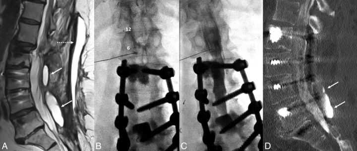
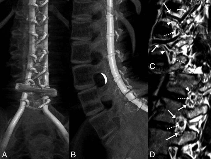
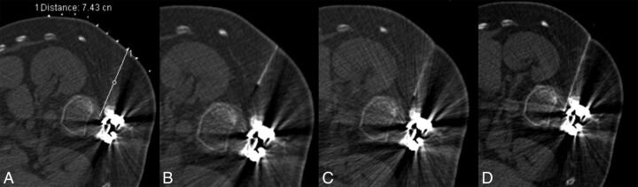
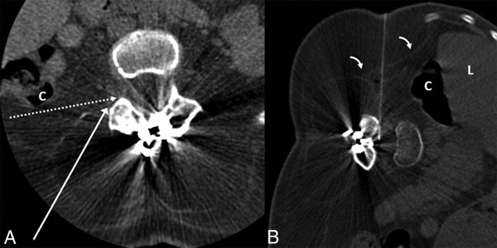
Interlaminar lumbar puncture and cervical puncture may not be ideal in all circumstances. Recently, we have used a transforaminal approach in selected situations. Between May 2016 and December 2017, twenty-six transforaminal lumbar punctures were performed in 9 patients (25 CT-guided, 1 fluoroscopy-guided). Seven had spinal muscular atrophy and were referred for intrathecal nusinersen administration. In 2, CT myelography was performed via transforaminal lumbar puncture. The lumbar posterior elements were completely fused in 8, and there was an overlying abscess in 1. The L1-2 level was used in 2; the L2-3 level, in 10; the L3-4 level, in 12; and the L4-5 level, in 2 procedures. Post-lumbar puncture headache was observed on 4 occasions, which resolved without blood patching. One patient felt heat and pain at the injection site that resolved spontaneously within hours. One patient had radicular pain that resolved with conservative treatment. Transforaminal lumbar puncture may become an effective alternative to classic interlaminar lumbar puncture or cervical puncture.


Figures

Fig 1.
Fig 1.
A 67-year-old woman with a history of multiple spinal fusions presented with newly worsening low back and radicular leg pain (case 1). Sagittal T2-weighted MR imaging (A) shows 2 paraspinous fluid collections (white arrows) within the deep paraspinal musculature and a midline subcutaneous fluid collection (dashed arrow) with rim enhancement on postcontrast series (not shown). B, Anteroposterior fluoroscopy image of myelography with TFLP. The needle tip is beyond the medial edge of the pedicle at the 5 o'clock position (relative to the pedicle). Note the position of the needle inferior to the expected location of the exiting nerve root and dorsal root ganglion. C, Oblique fluoroscopy image shows contrast extending into the intrathecal space after injection through the left L1–2 foramen. D, CT myelogram demonstrates the inferiorly located cystic collection filled with contrast, confirming a pseudomeningocele (arrows).
Fig 2.
Fig 2.
A and B, CT volumetric rendering of a patient with SMA2 demonstrates extensive posterior spinal fusion hardware and complete osseous interlaminar fusion without any access for a classic interlaminar LP. Note the widely patent neural foramina. The white crescent represents the target for TFLP. C and D, Sagittal 3D volumetric T2-weighted images of a healthy person obtained with 3T MR imaging. C, Image obtained slightly lateral to the neural foramen. D, Image obtained at the foramen. Flow voids of the lumbar arteries (arrowheads) and larger caliber lumbar veins (arrows) are seen in the anterior superior aspect of the foramen. A branching ascending lumbar vein is seen coursing toward a higher level neural foramen (curved arrow). More venous structures are seen in the inferior aspect of the foramen (arrows in D). Exiting nerve roots are shown within the center of the foramen (dashed arrows).
Fig 3.
Fig 3.
A 32-year-old man with SMA2 (case 5). A, Planning scout CT image with grid placement, obtained in the lateral decubitus position. B, Initial CT fluoroscopy image shows the needle more anteriorly oriented than desired. C, With basic needle manipulations, the needle is directed more posteriorly. D, Technically successful TFLP with the needle positioned immediately anterior to the facet.
Fig 4.
Fig 4.
A 36-year-old woman with SMA2 (case 6). A, Preprocedural CT obtained in supine position at the level of L3–4 demonstrates extensive muscle atrophy. The long white arrow shows the normal needle trajectory for a transforaminal epidural steroid injection. The dashed white arrow indicates the needle trajectory for TFLP. Note that while supine, the posterior margin of the ascending colon (C) is along the proposed needle trajectory. B, CT fluoroscopy image during TFLP obtained with the patient in the left lateral decubitus position. The needle is advanced into the thecal sac with an angle slightly >90°, just anterior to the facet. Note the anterior displacement of the liver (L) and ascending colon (C) in the decubitus position, providing a safer approach. Of note, the thin transversalis fascia (bent arrows) is well-visualized.

References

    1. Abel AS, Brace JR, McKinney AM, et al. . Practice patterns and opening pressure measurements using fluoroscopically guided lumbar puncture. AJNR Am J Neuroradiol 2012;33:823–25 10.3174/ajnr.A2876 - DOI - PMC - PubMed
    1. Cauley KA. Fluoroscopically guided lumbar puncture. AJR Am J Roentgenol 2015;205:W442–50 10.2214/AJR.14.14028 - DOI - PubMed
    1. Ozdoba C, Gralla J, Rieke A, et al. . Myelography in the age of MRI: why we do it, and how we do it. Radiol Res Pract 2011;2011:329017 10.1155/2011/329017 - DOI - PMC - PubMed
    1. Gibbs WN, Skalski MR, Kim PE, et al. . C1–2 puncture: a safe, efficacious, and potentially underused technique. Neurographics 2017;7:1–8 10.3174/ng.1170183 - DOI
    1. Pomerantz SR, Buchbinder B, Hirsch JA. Suboccipital puncture of the cisterna magna under CT-guidance with intravenous enhancement in order to circumvent anomalous course of posterior inferior cerebellar artery (PICA). In: Proceedings of the American Society of Spine Radiology Annual Symposium, San Juan, Puerto Rico February 24–27, 2005

LinkOut - more resources