. 2018 Mar 23;359(6382):1416-1421.
doi: 10.1126/science.aan1053.

Locally translated mTOR controls axonal local translation in nerve injury

Affiliations

Locally translated mTOR controls axonal local translation in nerve injury

Marco Terenzio et al. Science. .

Abstract

How is protein synthesis initiated locally in neurons? We found that mTOR (mechanistic target of rapamycin) was activated and then up-regulated in injured axons, owing to local translation of mTOR messenger RNA (mRNA). This mRNA was transported into axons by the cell size-regulating RNA-binding protein nucleolin. Furthermore, mTOR controlled local translation in injured axons. This included regulation of its own translation and that of retrograde injury signaling molecules such as importin β1 and STAT3 (signal transducer and activator of transcription 3). Deletion of the mTOR 3' untranslated region (3'UTR) in mice reduced mTOR in axons and decreased local translation after nerve injury. Both pharmacological inhibition of mTOR in axons and deletion of the mTOR 3'UTR decreased proprioceptive neuronal survival after nerve injury. Thus, mRNA localization enables spatiotemporal control of mTOR pathways regulating local translation and long-range intracellular signaling.


Conflict of interest statement

Competing interests: Alas, none.

Figures

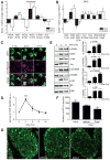
Fig. 1
Fig. 1. mTOR activation after nerve injury
(A) mTOR pathway phosphorylations significantly regulated by SN injury. n=3, * p < 0.05, *** p < 0.005, t-test. (B) As in A for L4/L5 DRG. n=3, * p < 0.05, t-test. (C) SN sections stained for the axonal marker NFH (green) and mTORS2448 (magenta), naive versus 3 and 6 hr after injury. Scale bar 5 μm. (D) Axonal mTORS2448 over time after injury, normalized to naive (n = 3, *** p < 0.005, one way ANOVA with Bonferroni’s post-test). (E) Immunoblots of phospho-EIF4b, Akt, S6K, and S6 and the corresponding total proteins in SN axoplasm over time after injury. Quantifications on the right (n = 4, * p < 0.05, ** p < 0.001, one way ANOVA with Bonferroni’s post-test). (F, G) SNs were injected with vehicle or Torin-1 prior to injury and L4 DRG were harvested 7 days later, serially sectioned at 20 μm intervals, and stained for NFH (green) to allow counting of proprioceptive neurons. Quantifications of NFH-positive neuron numbers per DRG are shown in F (n = 7, * p < 0.05, t-test), representative images are in G (scale bar 50 μm).
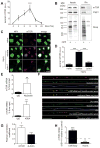
Fig. 2
Fig. 2. mTOR is locally translated after SN injury
(A) mTOR regulation over time after injury at the SN lesion site (n = 5, * p< 0.05, *** p<0.005, one way ANOVA with Bonferroni’s post-test). (B) Immunoblots reveal newly synthesized mTOR and Impβ1 from OPP treated rat SNs, confirming their local translation after injury. (C) Torin-1 (4 μM) effects on mTOR upregulation in sections from SN 4 hr ex vivo, stained for NFH (green) and mTOR (magenta). Scale bar 5 μm. (D) Quantification of axonal mTOR from C (n = 6, *** p < 0.005, one way ANOVA with Bonferroni’s post-test). (E) Quantification of mTOR transcript levels after pull down for Kif5A or nucleolin in axoplasm (n = 6), % from input, Mean ± SEM, ** denotes p < 0.01 (ratio paired t-test). (F) Representative epifluorescent images for co-localization of endogenous mTOR or β actin transcripts visualized by in situ hybridization (red) and nucleolin protein visualized by immunostaining (green). Axons were visualized by neurofilament immunostaining (blue). Scale bar 10 μm. (G) Pearsons correlation coefficient for mTOR mRNA colocalization with nucleolin (0.33 ± 0.04, n = 24) differs significantly from that of β actin mRNA colocalization with nucleolin (0.19 ± 0.04, n = 20). * denotes p < 0.05 (t-test). (H) Quantification of relative mTOR transcript levels in cell bodies and axons of neurons treated with AS1411 versus control aptamer, plotted as a fold change over control aptamer. 18S RNA served as an internal control. Mean ± SEM, n=3, * denotes p < 0.05 (unpaired two sample t-test).
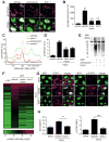
Fig. 3
Fig. 3. mTOR regulates axonal local translation after SN injury
(A) SN segments 2 hr ex vivo with anisomycin (200 μg/ml), torin-1 (4 μM) or vehicle, followed by 1 hr puromycin (100 μg/ml), sectioned and stained for NFH (green) and puromycin (magenta). Scale bar 5 μm. (B) Quantification of axonal puromycin in experiment described in A (n = 5, *** p < 0.001, one way ANOVA with Bonferroni’s post-test). (C) Representative runs of puromycinylated proteins in SN axoplasm from experiment described in A analysed by capillary immunoelectrophoresis. (D) Quantification of C (n=4, * p< 0.05, ** p<0.01, ANOVA with Bonferroni post-test). (E) SA-HRP immunoblots of OPP-biotin labeled axoplasm samples prior to MS. (F) Heat map of OPP-biotin labeled protein candidates identified by MS. (G) SN segments 4 hr ex vivo with Torin-1 (4 μM) or vehicle (DMSO), sectioned and stained for NFH (green) and STAT3 or phosphorylated STAT3 (pSTAT3, both magenta). Scale bars 5 μm. (H) Quantification of axonal STAT3 and p-STAT3 for the experiment described in G (n = 4, ** p < 0.01, *** p < 0.005, one way ANOVA with Bonferroni’s post-test).
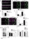
Fig. 4
Fig. 4. Effects of mTOR 3′UTR deletion
(A) Representative, exposure-matched confocal images of Stellaris FISH for mTOR mRNA and neurofilament (NF) immunostaining from SN sections of wild type and mTOR 3′UTR−/− mice. Upper panels show single optical planes for merged NFH and mTOR channels. Lower panels show single optical planes of mTOR mRNA pixels that overlap with NFH and were projected to a separate channel as ‘axon only’ mTOR signals. Scale bar 10 μm. (B) Quantification of A reveals approximately 50% reduction in axonal mTOR in the 3′UTR−/− mice (n = 4, ** p < 0.01, unpaired t-test). (C) SN segments from the indicated genotypes 4 hr ex vivo, sectioned and stained for NFH (green) and mTOR (magenta). Scale bar 5 μm. (D) Quantification of C (n = 3, * p < 0.05, one way ANOVA with Bonferroni’s post-test). (E) SN segments from the indicated genotypes 2 hr ex vivo with anisomycin (200 μg/ml) or vehicle, followed by 1 hr puromycin (100 μg/ml), then sectioned and stained as indicated. Scale bar 5 μm. (F) Quantification of E (n = 3, ** p < 0.01, one way ANOVA with Bonferroni’s post-test). (G) SN segments from wild type and mTOR 3′UTR/ mice not injected, injected with vehicle and injected with 350 ng mTOR protein were incubated in DMEM 2 hr ex vivo, followed by 1 hr puromycin (100 μg/ml) treatment. A representative pseudo-blot of puromycinylated proteins in SN axoplasm analysed by capillary immunoelectrophoresis is shown. (H) Quantification of G (n=4, *** p< 0.005, one way ANOVA with Bonferroni’s post-test). (I) SNs from wild type and mTOR 3′UTR−/− mice were injected with either vehicle or 350 ng of mTOR protein concomitant with crush injury. L4 DRGs connected to the injured sciatic were harvested 7 days after injury, serially sectioned at 20 μm intervals and stained for the proprioceptive marker NFH. Naive L4 DRG were also processed as a reference. Number of NFH positive neurons per DRG (n = 4, * p < 0.05, ** p < 0.01, one way ANOVA with Tukey’s post-test).

Comment in

Similar articles

Cited by

References

    1. Rangaraju V, Tom Dieck S, Schuman EM. Local translation in neuronal compartments: how local is local? EMBO Rep. 2017;18:693–711. - PMC - PubMed
    1. Terenzio M, Schiavo G, Fainzilber M. Compartmentalized signaling in neurons: from cell biology to neuroscience. Neuron. 2017;96:667–679. - PubMed
    1. Hanz S, et al. Axoplasmic importins enable retrograde injury signaling in lesioned nerve. Neuron. 2003;40:1095–1104. - PubMed
    1. Perry RB, et al. Subcellular knockout of importin beta1 perturbs axonal retrograde signaling. Neuron. 2012;75:294–305. - PMC - PubMed
    1. Zheng JQ, et al. A functional role for intra-axonal protein synthesis during axonal regeneration from adult sensory neurons. J Neurosci. 2001;21:9291–9303. - PMC - PubMed

Publication types