Long-Term Morphological Changes of Symptomatic Lacunar Infarcts and Surrounding White Matter on Structural Magnetic Resonance Imaging
- PMID: 29567763
- PMCID: PMC5916475
- DOI: 10.1161/STROKEAHA.117.020495
Long-Term Morphological Changes of Symptomatic Lacunar Infarcts and Surrounding White Matter on Structural Magnetic Resonance Imaging
Abstract
Background and purpose: Insights into evolution of cerebral small vessel disease on neuroimaging might advance knowledge of the natural disease course. Data on evolution of sporadic symptomatic lacunar infarcts are limited. We investigated long-term changes of symptomatic lacunar infarcts and surrounding white matter on structural magnetic resonance imaging.
Methods: From 2 nonoverlapping, single-center, prospective observational stroke studies, we selected patients presenting with lacunar stroke symptoms with a recent small subcortical (lacunar) infarct on baseline structural magnetic resonance imaging and with follow-up magnetic resonance imaging available at 1 to 5 years. We assessed changes in imaging characteristics of symptomatic lacunar infarcts and surrounding white matter.
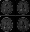
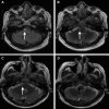
Results: We included 79 patients of whom 32 (41%) had complete and 40 (51%) had partial cavitation of the index lesion at median follow-up of 403 (range, 315-1781) days. In 42 of 79 (53%) patients, we observed a new white matter hyperintensity adjacent to the index infarct, either superior (white matter hyperintensity cap, n=17), inferior (white matter hyperintensity track, n=13), or both (n=12).
Conclusions: Half of the sporadic symptomatic lacunar infarcts developed secondary changes in superior and inferior white matter. These white matter hyperintensity caps and tracks may reflect another aspect of cerebral small vessel-related disease progression. The clinical and prognostic values remain to be determined.
Keywords: Wallerian degeneration; cerebral small vessel diseases; follow-up studies; lacunar; stroke.
© 2018 The Authors.
Figures


Comment in
-
Letter by Ryan et al Regarding Article, "Long-Term Morphological Changes of Symptomatic Lacunar Infarcts and Surrounding White Matter on Structural Magnetic Resonance Imaging".Stroke. 2018 Jul;49(7):e268. doi: 10.1161/STROKEAHA.118.021653. Epub 2018 Jun 18. Stroke. 2018. PMID: 29915119 No abstract available.
-
Response by Loos et al to Letter Regarding Article, "Long-Term Morphological Changes of Symptomatic Lacunar Infarcts and Surrounding White Matter on Structural Magnetic Resonance Imaging".Stroke. 2018 Jul;49(7):e269. doi: 10.1161/STROKEAHA.118.021876. Epub 2018 Jun 18. Stroke. 2018. PMID: 29915121 Free PMC article. No abstract available.
References
-
- Wardlaw JM, Smith EE, Biessels GJ, Cordonnier C, Fazekas F, Frayne R, et al. Standards for Reporting Vascular Changes on Neuroimaging (STRIVE v1) Neuroimaging standards for research into small vessel disease and its contribution to ageing and neurodegeneration. Lancet Neurol. 2013;12:822–838. doi: 10.1016/S1474-4422(13)70124-8. - PMC - PubMed
-
- Potter GM, Doubal FN, Jackson CA, Chappell FM, Sudlow CL, Dennis MS, et al. Counting cavitating lacunes underestimates the burden of lacunar infarction. Stroke. 2010;41:267–272. doi: 10.1161/STROKEAHA.109.566307. - PubMed
-
- Koch S, McClendon MS, Bhatia R. Imaging evolution of acute lacunar infarction: leukoariosis or lacune? Neurology. 2011;77:1091–1095. doi: 10.1212/WNL.0b013e31822e1470. - PubMed
-
- Loos CM, Staals J, Wardlaw JM, van Oostenbrugge RJ. Cavitation of deep lacunar infarcts in patients with first-ever lacunar stroke: a 2-year follow-up study with MR. Stroke. 2012;43:2245–2247. doi: 10.1161/STROKEAHA.112.660076. - PubMed
Publication types
MeSH terms
Grants and funding
LinkOut - more resources
Full Text Sources
Other Literature Sources
Miscellaneous

