. 2018 Apr;8(4):403-416.
doi: 10.1158/2159-8290.CD-17-1134. Epub 2018 Mar 22.

The Pancreatic Cancer Microbiome Promotes Oncogenesis by Induction of Innate and Adaptive Immune Suppression

Affiliations

The Pancreatic Cancer Microbiome Promotes Oncogenesis by Induction of Innate and Adaptive Immune Suppression

Smruti Pushalkar et al. Cancer Discov. 2018 Apr.

Erratum in

Abstract

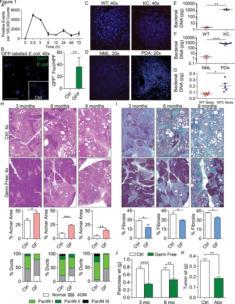
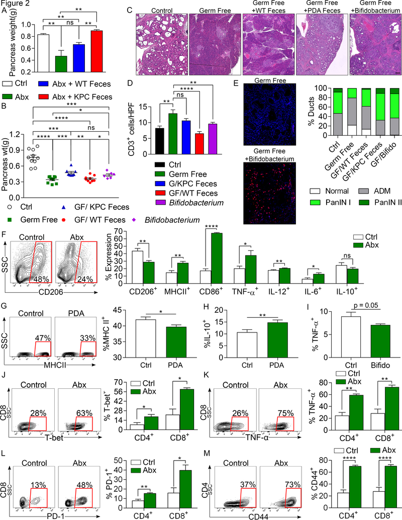
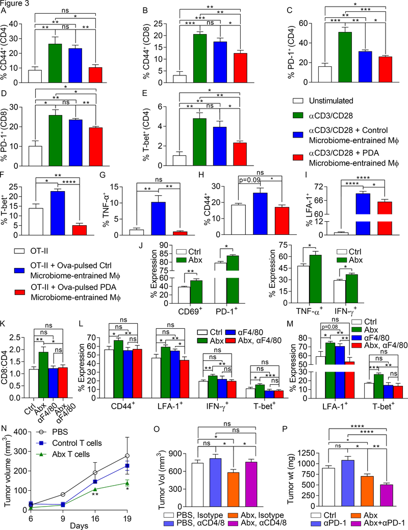
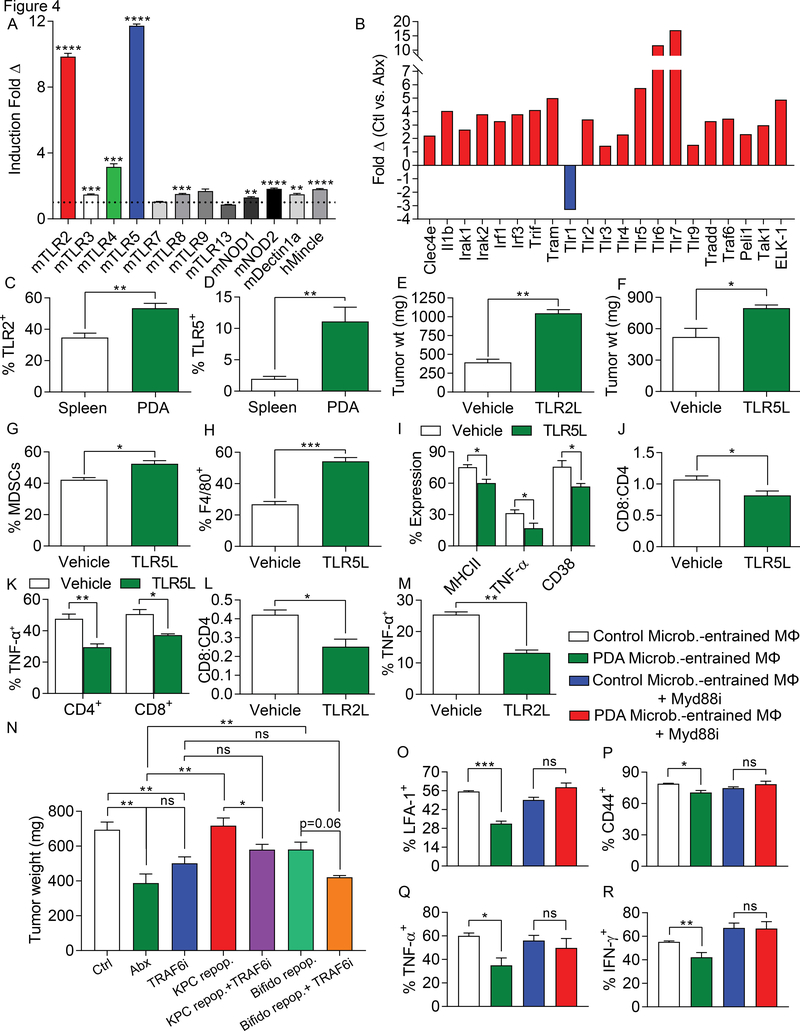
We found that the cancerous pancreas harbors a markedly more abundant microbiome compared with normal pancreas in both mice and humans, and select bacteria are differentially increased in the tumorous pancreas compared with gut. Ablation of the microbiome protects against preinvasive and invasive pancreatic ductal adenocarcinoma (PDA), whereas transfer of bacteria from PDA-bearing hosts, but not controls, reverses tumor protection. Bacterial ablation was associated with immunogenic reprogramming of the PDA tumor microenvironment, including a reduction in myeloid-derived suppressor cells and an increase in M1 macrophage differentiation, promoting TH1 differentiation of CD4+ T cells and CD8+ T-cell activation. Bacterial ablation also enabled efficacy for checkpoint-targeted immunotherapy by upregulating PD-1 expression. Mechanistically, the PDA microbiome generated a tolerogenic immune program by differentially activating select Toll-like receptors in monocytic cells. These data suggest that endogenous microbiota promote the crippling immune-suppression characteristic of PDA and that the microbiome has potential as a therapeutic target in the modulation of disease progression.Significance: We found that a distinct and abundant microbiome drives suppressive monocytic cellular differentiation in pancreatic cancer via selective Toll-like receptor ligation leading to T-cell anergy. Targeting the microbiome protects against oncogenesis, reverses intratumoral immune tolerance, and enables efficacy for checkpoint-based immunotherapy. These data have implications for understanding immune suppression in pancreatic cancer and its reversal in the clinic. Cancer Discov; 8(4); 403-16. ©2018 AACR.See related commentary by Riquelme et al., p. 386This article is highlighted in the In This Issue feature, p. 371.


Conflict of interest statement

Competing financial interests

None of the authors have any competing financial interests to declare.

The authors declare no potential conflicts of interest.

Figures

Figure 1.
Figure 1.. The tumorous pancreas has an abundant microbiome and its ablation is protective against pancreatic disease progression.
(A) WT mice were administered CFSE-labeled E. faecalis (2.5×10⁸ CFU) via oral gavage. Pancreata were harvested and digested at the indicated timed intervals and tested for the presence of these bacteria (n=3 mice/time point). This experiment was repeated twice with similar results. (B) WT mice were administered GFP-labeled E. coli (2.5×10⁸ CFU) via oral gavage. Pancreata were harvested at 6h and the number of GFP+ foci was determined by immune fluorescence microscopy compared to control. This experiment was repeated twice (n=3; **p<0.01; scale bar =50μm). (C) The abundance of intra-pancreatic bacteria was compared in 3-month-old WT and KC mice by FISH (n=5/group). Representative images are shown. This experiment was repeated twice. (D) The abundance of intra-pancreatic bacteria was compared in healthy individuals and age/gender/BMI matched PDA patients by FISH (n=5/group). Representative images are shown. (E) Bacterial DNA content was compared in WT and KC mice using qPCR. Each dot represents data from a single mouse pancreas. This was repeated three times (**p<0.01). (F) Bacterial DNA content was compared in healthy individuals (NML) and age/gender/BMI matched PDA patients using qPCR. Each dot represents data from a single human pancreas (****p<0.0001). (G) 8-week old WT mice were treated with an ablative oral antibiotic regimen. 3 weeks after treatment, mice were repopulated using fecal bacteria from either 3 month-old WT or KPC mice. Bacterial colonization of the pancreas was analyzed by qPCR 2 weeks after repopulation. This experiment was repeated twice (n=5/group; *p<0.05). (H-J) Control and germ-free KC mice were sacrificed at 3, 6, or 9 months of life. Representative (H) H&E- and (I) trichrome-stained sections are shown. The percentage of ducts exhibiting normal morphology, acinoductal metaplasia (ADM), or graded PanIN lesions were determined based on H&E staining. The fraction of fibrotic area per pancreas was calculated based on trichrome staining (scale bars = 200μm). (J) Pancreatic weights were recorded at 3 or 6 months of life (n=10/group; *p<0.05, **p<0.01, ***p<0.001, ****p<0.0001). (K) WT mice were treated with an ablative oral antibiotic regimen and then orthotopically inoculated with KPC-derived PDA cells. Animals were sacrificed at 3 weeks and tumor weights were recorded (n=4/group; **p<0.01). This experiment was repeated more than 5 times with similar results.
Figure 2.
Figure 2.. The microbiome in PDA-bearing hosts promotes tumor-progression and intra-tumoral immune suppression.
(A) KC mice treated with an ablative oral antibiotic regimen for 8 weeks were repopulated with i) feces from 3-month old WT mice, ii) feces from 3 month-old KPC mice, or iii) sham-repopulated (vehicle only). Mice were sacrificed 8 weeks later and pancreas weights from each cohort were compared to each other and to age-matched control KC mice that were not treated with antibiotics (n=3–4/group). This experiment was repeated three times. (B-D) The gut microbiome of germ-free 6-week-old KC mice were repopulated with feces from 3-month-old WT or KPC mice, B. pseudolongum, or sham-repopulated. Mice were sacrificed 8 weeks later. (B) Tumor weights were measured. Each point represents data from a single mouse. (C) Representative H&E-stained sections of pancreata are shown compared with age-matched non-germ free controls (scale bar =100μm). Ductal histology was quantified. (D) CD3+ T cell infiltration was determined by IHC. All repopulation experiments were repeated 3 times. (E) The gut microbiome of germ-free 6-week-old KC mice were repopulated with B. pseudolongum or sham-repopulated (n=5/group). Colonization of pancreata with B. pseudolongum was confirmed using FISH at 8 weeks. This experiment was repeated twice. (F) Control and oral antibiotic-treated WT mice were orthotopically implanted with KPC-derived tumor cells. Gr1CD11b+F4/80+ macrophages were gated and assessed for expression of CD206, MHC II, CD86, TNF-α, IL-12, IL-6, and IL-10 (n=5/group). Macrophage profiling experiments were repeated more than 5 times (G, H) Splenic macrophages from untreated mice were harvested and cultured in vitro with cell-free extract from gut bacteria of control or KC mice. After 24h, macrophages were analyzed for expression of (G) MHC II and (H) IL-10 (n=18/group). (I) Splenic macrophages were cultured in vitro with cell-free extract from B. pseudolongum or with PBS. After 24h, macrophages were analyzed for expression of TNF-α. Macrophage polarization experiments were repeated 3 times in replicates of 5. (J-M) Control and oral antibiotic-treated WT mice were orthotopically implanted with KPC-derived tumor cells. CD4+ and CD8+ T cells were gated and tested for expression of (J) T-bet, (K) TNF-α, (L) PD-1, and (M) CD44. Representative contour plots and quantitative data are shown. Immune-phenotyping experiments were repeated more than 5 times (n=5/group; *p<0.05, **p<0.01, ***p<0.001, ****p<0.0001).
Figure 3.
Figure 3.. The PDA microbiome promotes macrophage-mediated suppression of T cell immunity.
(A-E) Naïve splenic CD4+ and CD8+ T cells from WT mice were activated using CD3/CD28 co-ligation, either alone or in the presence of splenic macrophages that had been treated overnight with cell-free extract from gut bacteria derived from 3-month-old WT mice or KC mice. CD4+ and CD8+ T cell activation, respectively, were determined by expression of (A, B) CD44 and (C, D) PD-1. (E) CD4+ T cell differentiation was further evaluated by expression of T-bet. This experiment was repeated more than 5 times using 3–5 replicates per group. (F-I) Splenic macrophages that had been treated overnight with cell-free extract from gut bacteria derived from 3-month-old WT mice or KC mice were pulsed with Ova323–339 peptide and used to stimulate CD4+ OT-II T cells. T cell activation at 96h was determined by expression of (F) T-bet, (G) TNF-α, (H) CD44, and (I) LFA-1. This experiment was repeated 4 times using 4–5 replicates per group. (J) Control and oral antibiotic-treated WT mice bearing orthotopic KPC-derived tumor cells were sacrificed at 21 days. TAMs were FACS-sorted, loaded with Ova257–264 peptide, and used to stimulate Ova-restricted CD8+ OT-I T cells. T cell activation was determined by expression of TNF-α, IFN-γ, CD69, and PD-1. This experiment was repeated 3 times (n=5/group). (K-M) Cohorts of orthotopic PDA-bearing mice treated with oral PBS or ablative antibiotics were serially administered neutralizing αF480 or isotype control (n=10/group). Mice were sacrificed at 21 days and tumor-infiltrating T cells were analyzed for (K) the CD8:CD4 ratio, (L) CD4+ T cell expression of CD44, LFA-1, IFN-γ, and T-bet, and (M) CD8+ T cell expression of LFA-1 and T-bet. (N) PDA-infiltrating T cells from orthotopic KPC tumor-bearing mice that had been treated with an ablative oral antibiotic regimen or sham-treated were harvested on day 21 by FACS, mixed with FC1242 cells in a 1:10 ratio, and subcutaneously implanted in the flank of recipient mice. Additional controls received FC1242 cells alone. Tumor volumes were measured at serial intervals. This experiment was repeated 3 times (n=4/group). (O) Cohorts of orthotopic PDA-bearing mice treated with oral PBS or ablative antibiotics were serially administered neutralizing αCD4 and αCD8 mAbs or isotype control. Mice were sacrificed at 21 days and pancreatic tumors were weighed (n=8–9/group). T cell depletion experiments were performed more than 3 times in orthotopic PDA-bearing mice. (P) WT mice were treated with vehicle (n=9), αPD-1 (n=16), an ablative oral antibiotic regimen (n=6), or both (n=9). Mice were challenged with orthotopic KPC tumor and sacrificed at 3 weeks. Treatments were started before tumor implantation and continued until the time of sacrifice. This experiment was repeated 4 times (n=10/group; *p<0.05, **p<0.01, ***p<0.001, ****p<0.0001).
Figure 4.
Figure 4.. The PDA microbiome induces immune suppression via differential TLR activation.
(A) Cell-free extract from gut bacteria derived from 3 months old WT or KC mice (n=3) were tested for activation of a diverse array of PRR-specific HEK293 reporter cell lines. (B) Orthotopic KPC tumors were harvested on day 21 from control and oral antibiotic-treated WT mice. PRR-related gene expression in PDA was determined using a PCR array and performed in duplicate. Data indicates fold change in gene expression for control compared to antibiotic-treated groups. This array was repeated twice. (C) Expression of TLR2 and (D) TLR5 were tested in spleen and PDA-infiltrating macrophages from orthotopic KPC tumors (n=5). These experiments were repeated twice. (E) WT mice were orthotopically implanted with KPC-derived tumor cells and serially treated with TLR2 (Pam3CSK4) or (F) TLR5 (Flagellin) ligand or vehicle. Tumor growth was determined at 3 weeks (n=3–5/group). These experiments were repeated twice. (G-K) WT mice were orthotopically implanted with KPC-derived tumor cells and serially treated with TLR5 ligand. Tumors were harvested at 3 weeks and (G) the fraction of Gr1+CD11b+ MDSC and (H) F4/80+Gr1CD11b+ TAM infiltration was determined by flow cytometry. (I) Expression of MHC II, TNF-α, and CD38 on TAMs were determined. (J) The CD8/CD4 T cell ratio was determined as was (K) TNF-α expression on CD4+ and CD8+ T cells (n=3–5/group). (L, M) WT mice were orthotopically implanted with KPC-derived tumor cells and serially treated with TLR2 ligand. Tumors were harvested at 3 weeks and (L) the CD8/CD4 T cell ratio and (M) TAMs expression of TNF-α were determined. These experiments were repeated twice (n=3–5/group). (N) WT mice pre-treated with an ablative oral antibiotic regimen or vehicle for 6 weeks were repopulated with i) feces from 3 month-old KPC mice (n=12), ii) B. pseudolongum (n=6), or iii) sham-repopulated (n=4). Mice were challenged with orthotopic KPC cells. Cohorts were additionally treated serially with a TRAF6 inhibitor or control. Treatments were started at the time of tumor implantation and continued until sacrifice at 21 days. Quantitative analysis of tumor weights are shown. (O-R) Splenic macrophages were entrained with extract from the gut microbiome of either WT or KC mice in the context of MyD88 inhibition or control. Macrophages were then used in αCD3/αCD28-based T cell stimulation assays. CD4+ T cell activation was determined by expression of (O) LFA-1, (P) CD44, (Q) TNF-α, and (R) IFN-γ. This experiment was repeated five times in 3–4 replicates per group with similar results (*p<0.05, **p<0.01, ***p<0.001, ****p<0.0001).

Comment in

References

    1. Siegel RL, Miller KD and Jemal A (2016) Cancer statistics, 2016. CA: a cancer journal for clinicians, 66, 7–30. - PubMed
    1. Zambirinis CP, Levie E, Nguy S, Avanzi A, Barilla R, Xu Y, Seifert L, Daley D, Greco SH, Deutsch M et al. (2015) TLR9 ligation in pancreatic stellate cells promotes tumorigenesis. The Journal of experimental medicine, 212, 2077–2094. - PMC - PubMed
    1. Ibrahim J, Nguyen AH, Rehman A, Ochi A, Jamal M, Graffeo CS, Henning JR, Zambirinis CP, Fallon NC, Barilla R et al. (2012) Dendritic cell populations with different concentrations of lipid regulate tolerance and immunity in mouse and human liver. Gastroenterology, 143, 1061–1072. - PMC - PubMed
    1. Ochi A, Nguyen AH, Bedrosian AS, Mushlin HM, Zarbakhsh S, Barilla R, Zambirinis CP, Fallon NC, Rehman A, Pylayeva-Gupta Y et al. (2012) MyD88 inhibition amplifies dendritic cell capacity to promote pancreatic carcinogenesis via Th2 cells. The Journal of experimental medicine, 209, 1671–1687. - PMC - PubMed
    1. Zambirinis CP, Ochi A, Barilla R, Greco S, Deutsch M and Miller G (2013) Induction of TRIF- or MYD88-dependent pathways perturbs cell cycle regulation in pancreatic cancer. Cell cycle, 12, 1153–1154. - PMC - PubMed

Publication types

Substances