. 2018 Apr 15;78(8):2052-2064.
doi: 10.1158/0008-5472.CAN-17-2994. Epub 2018 Mar 22.

Targeting Brain-Adaptive Cancer Stem Cells Prohibits Brain Metastatic Colonization of Triple-Negative Breast Cancer

Affiliations

Targeting Brain-Adaptive Cancer Stem Cells Prohibits Brain Metastatic Colonization of Triple-Negative Breast Cancer

Ding Ren et al. Cancer Res. .

Abstract

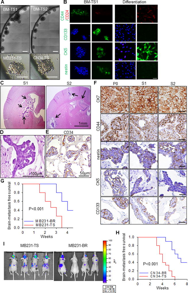
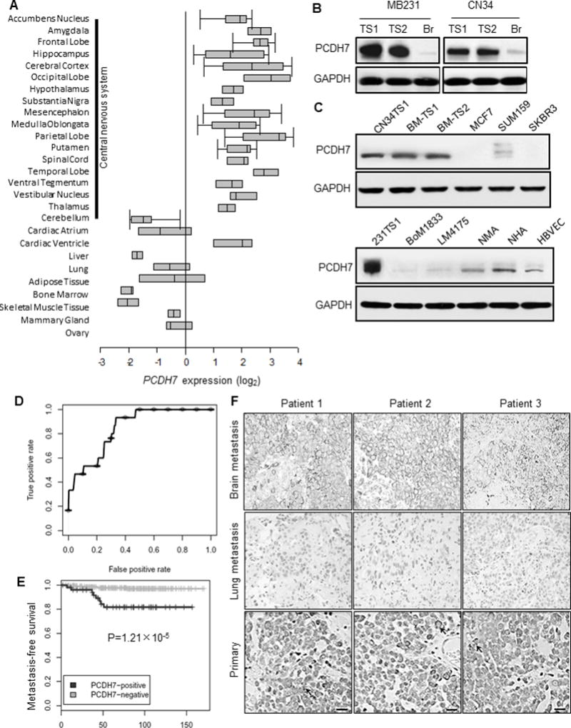
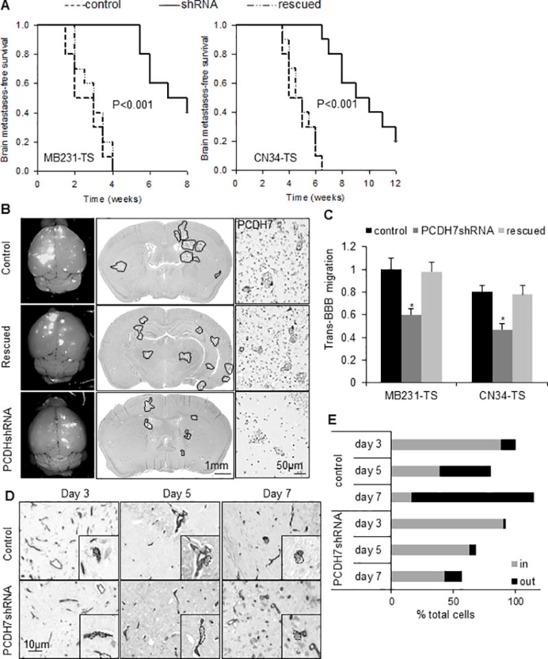
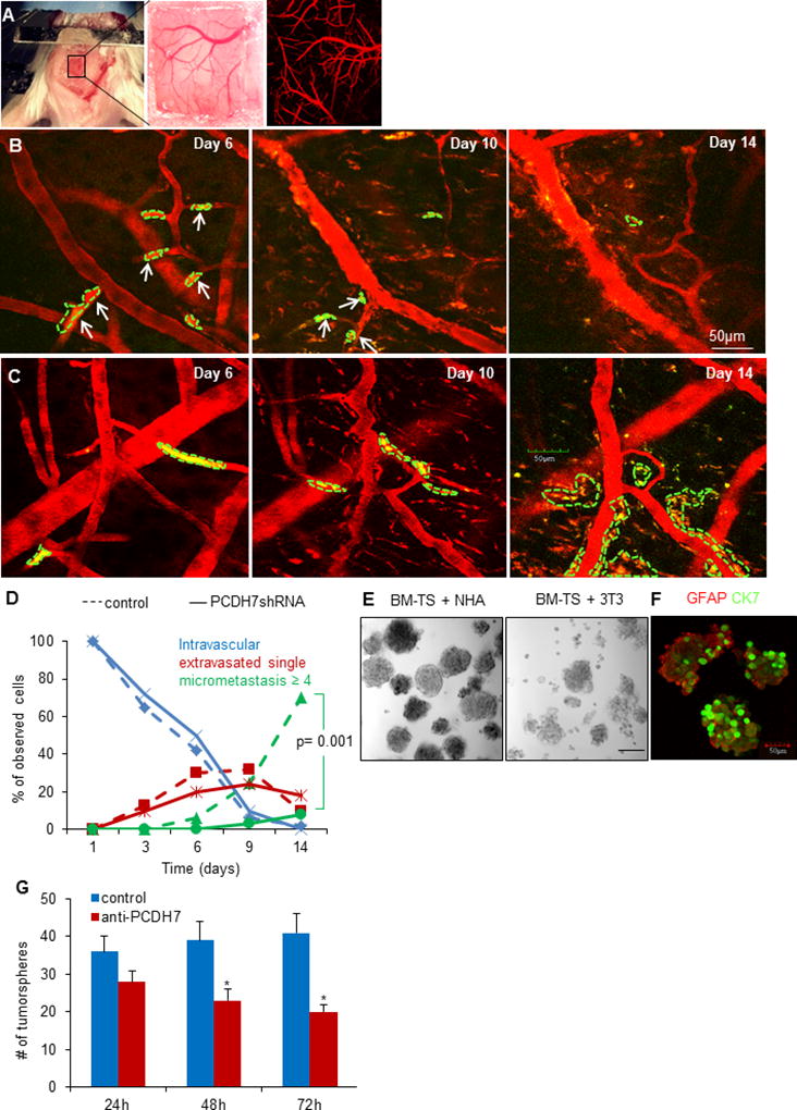
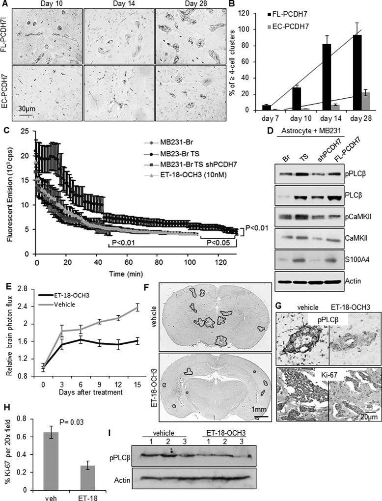
Triple-negative breast cancer (TNBC) exhibits more traits possessed by cancer stem cells (CSC) than other breast cancer subtypes and is more likely to develop brain metastases. TNBC patients usually have shorter survival time after diagnosis of brain metastasis, suggesting an innate ability of TNBC tumor cells in adapting to the brain. In this study, we establish novel animal models to investigate early tumor adaptation in brain metastases by introducing both patient-derived and cell line-derived CSC-enriched brain metastasis tumorsphere cells into mice. We discovered astrocyte-involved tumor activation of protocadherin 7 (PCDH7)-PLCβ-Ca2+-CaMKII/S100A4 signaling as a mediator of brain metastatic tumor outgrowth. We further identified and evaluated the efficacy of a known drug, the selective PLC inhibitor edelfosine, in suppressing the PCDH7 signaling pathway to prohibit brain metastases in the animal models. The results of this study reveal a novel signaling pathway for brain metastases in TNBC and indicate a promising strategy of metastatic breast cancer prevention and treatment by targeting organ-adaptive cancer stem cells.Significance: These findings identify a compound to block adaptive signaling between cancer stem cells and brain astrocytes. Cancer Res; 78(8); 2052-64. ©2018 AACR.


Conflict of interest statement

Disclosure of Potential Conflicts of Interest: No potential conflicts of interest were disclosed.

Figures

Fig. 1
Fig. 1. Brain metastasis models derived from brain metastasis-derived tumorspheres
A. Bright-field images of representative tumorspheres cultured from fresh brain metastases tissue from two TNBC patient undergoing neurosurgical resection, i.e., BM-TS1 and BM-TS2, and brain seeking cell lines, i.e., MDA-MB231-TS and CN34-TS. Scale bar is 50µm. B. The BM-TS1 tumorspheres were stained with antibodies to antibodies to CD44, CD24, CD133, CK5 and nestin. For differentiation, BM-TSs were disrupted and the cells plated onto glass coverslips in medium supplemented with 5% fetal calf serum. After 10 days of adherent culture, the cells were stained with the same antibodies. scale bar: 20µm. C. Representative H.E. staining images showing the intraparenchymal multifocal S1 and S2 brain metastatic tumors in mice. Mice were injected with 1.75×10⁵ BM-TS dissociated cells through the left ventricle. D. The cytoarchitecture of the S1/S2 tumor. E. The vessel cooption growth pattern of the S1/S2 tumors. F. The expression pattern of CK7, CD44, nestin, CK5 and CD133 in the original patient P0 brain metastatic tumor and serially passaged S1 and S2 tumors. G–H. Kaplan-Meier survival curves for brain metastasis-free survival of tumorsphere derived models (MB231-TS, CN34-TS) in comparing with brain seeking cell line derived models (MB231-Br, CN34-Br). P values were determined by log rank test. N=10 in each group. I. The representative bioluminescent imaging of the brain metastases in MB231-TS and MB-231-Br models.
Fig. 2
Fig. 2. High expression of PCDH7 in brain metastasis
A. Oncomine database analysis of PCDH7 expression in normal human tissue (Neurogenetics 2006 7:67–80). B. Western Blot analysis of PCDH7 in independent brain metastasis-derived tumorspheres (TS1 and TS2) and corresponding brain-seeking (Br) cell lines. C. Protein expression of PCDH7 in TNBC patient brain metastasis tumorspheres (BM-TS1 and 2) and various cell models. MCF7, SUM159, SKBR3: human breast cancer cell lines; BoM1833: MB231 bone-seeking cell line; LM4175: MB231 lung seeking cell line; NMA: primary normal mouse astrocytes; NHA: normal human astrocytes; HBVEC: human brain microvascular endothelial cells. D. Receiver Operating Characteristic (ROC) curve for PCDH7 expression in primary breast tumor samples of brain metastatic patients using the combined 368 microarray data (MSK-82 and EMC-286 cohort). E. Kaplan–Meier curves showing the brain metastasis-free survival of patients with positive or negative PCDH7 expression in the combined cohort of 368 breast cancer patients, P=1.21×10⁻⁵ determined by log rank test. F. Representative PCDH7 immunohistochemistry staining of matched patient tissue sections of brain metastasis, lung metastasis and primary breast tumors. Scale bar: 20µm.
Fig. 3
Fig. 3. Functional roles of PCDH7 in brain metastasis
A. Kaplan-Meier curves for brain metastasis-free survival of mice injected with tumorsphere cells derived from MB231-Br (left) or CN34=Br (right) models expressing the shRNA targeting PCDH7, the control shRNA or the PCDH7 rescued. n=10 mice per group. P value was determined using log rank test. B. Representative images showing metastatic lesions in mouse whole brain (left column), H&E stained brain section (middle column), and PCDH7 immuno-reactivities in the brain metastases (right column). It’s noted that high expression of PCDH7 in both tumor cells and surrounding astrocytes in control group was diminished by PCDH7shRNA. n=5 mice per group. C. Transmigration of the indicated cells through the in vitro BBB system. * P<0.05, vs. control shRNA. D. Representative images showing intravascular and extravasated tumor cells in mouse brain sections. MB231-TS cells were visualized on 10µm-thick brain sections by anti-human CD44 and blood vessels by anti-mouse CD34 using immunohistochemistry. Black rectangles are higher magnification single cell images. CD44+ tumor cells were segmented by dash lines. E. Percentage of cancer cells located inside (in) vs. outside (out) blood vessels at indicated days after intracardiac tumor cell injection. Intravascular and extravasated tumor cells were counted in every fourth section throughout the entire mouse brain. Data was relative to “control group day 3”. It’s noted that the % total cells > 100% is due to the cell proliferation, and the % total cells < 100% is due to cell disappear. n=3 mice per each time point.
Fig. 4
Fig. 4. PCDH7 mediates the interaction between cancer cells and astrocytes to promote tumor colonization
A. A thinned-skull window with perfused brain vessels (Texas-red dextran, red). B. Intravital two-photon microscopy images showing tumor cell arrest and extravasation in mouse brain at day 6, and disappeared at day 10 and day 14 (indicated as solid white arrows) for PCDH7shRNA MB231-TS cells, while the control shRNA cells formed colonization at day 14 (C). It’s noted that the blood vessels became tortuous and leaky after day 6 and remodeled at day 14. D. Extravasation and colonization of the indicated cells in mouse brain over time. n=4 mice per group. E. Representative bright-filed images of tumorspheres at passage 2 when co-culturing BM-TS cells with NHA or normal fibroblasts 3T3 cells. F. Representative immunofluorescent images showing the heteotypic spheres formed by cancer cells (stained for CK7) and astrocytes (stained for GFAP). G. Effects of PCDH7 neutralization by anti-PCDH7 antibody (1:100) on tumorsphere formation (diameter >50µm) in the dissociated BM-TS cells and NHAs co-culturing system. * P<0.05, vs. control.
Fig. 5
Fig. 5. Tumor-astrocyte interaction activates PCDH7-PLCβ-Ca2+ signaling to promote colonization
A. Representative immunohistochemistry images of metastatic colonies in mouse brain sections with intra-cardiac injection of indicated cells. Tumor cells were visualized with anti-human CD44 antibody and blood vessels were visualized with anti-mouse CD34 antibody. B. Percent of ≥ 4-cell clusters located outside vessels at indicated days after tumor cell injection (n=3 mice/time point/cell line). C. Baseline level of cytoplasmic Ca2+ in the indicated tumor cells measure by ratiometric emission of calcium-saturated Asante of the tumor cells when co-culturing with astrocytes. D. Signal activity of PLCβ, CaMKII and S100A in the indicated MB231 cells when co-culturing with normal human astrocytes. E. In vivo effects of PLC selective inhibitor ET-18-OCH3 (i.p., 30mg/kg, once daily) on brain metastatic tumor growth. n=10 per group. P=0.04, determined by ANOVA post-hoc test. F. Representative whole-brain sections showing macro-metastatic lesions of ET-18-OCH3 or vehicle-treated mice at the end of the treatment. G. Representative immunostained images of pPLCβ1 and Ki-67 in brain sections. H. Quantification of the % of Ki-67-positive tumor cells in the brain sections. P=0.03, determined by student’s t-test. I. Western blot of pPLCβ1 expression in mouse brain lysates. N=3 per group.

Similar articles

Cited by

References

    1. Qiu J, Xue X, Hu C, Xu H, Kou D, Li R, et al. Comparison of Clinicopathological Features and Prognosis in Triple-Negative and Non-Triple Negative Breast Cancer. J Cancer. 2016;7(2):167–73. - PMC - PubMed
    1. Owonikoko TK, Arbiser J, Zelnak A, Shu HK, Shim H, Robin AM, et al. Current approaches to the treatment of metastatic brain tumours. Nat Rev Clin Oncol. 2014;11(4):203–22. - PMC - PubMed
    1. Leone JP, Leone BA. Breast cancer brain metastases: the last frontier. Exp Hematol Oncol. 2015;4:33. - PMC - PubMed
    1. Niikura N, Hayashi N, Masuda N, Takashima S, Nakamura R, Watanabe K, et al. Treatment outcomes and prognostic factors for patients with brain metastases from breast cancer of each subtype: a multicenter retrospective analysis. Breast Cancer Res Treat. 2014;147(1):103–12. - PubMed
    1. Oskarsson T, Batlle E, Massague J. Metastatic stem cells: sources, niches, and vital pathways. Cell Stem Cell. 2014;14(3):306–21. - PMC - PubMed

Publication types

MeSH terms