. 2018 Mar 22;9(1):1184.
doi: 10.1038/s41467-018-03391-w.

Surface tension-assisted additive manufacturing

Affiliations

Surface tension-assisted additive manufacturing

Héloïse Ragelle et al. Nat Commun. .

Abstract

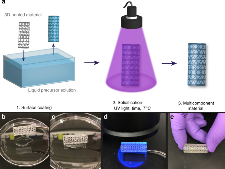
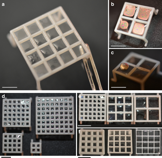
The proliferation of computer-aided design and additive manufacturing enables on-demand fabrication of complex, three-dimensional structures. However, combining the versatility of cell-laden hydrogels within the 3D printing process remains a challenge. Herein, we describe a facile and versatile method that integrates polymer networks (including hydrogels) with 3D-printed mechanical supports to fabricate multicomponent (bio)materials. The approach exploits surface tension to coat fenestrated surfaces with suspended liquid films that can be transformed into solid films. The operating parameters for the process are determined using a physical model, and complex geometric structures are successfully fabricated. We engineer, by tailoring the window geometry, scaffolds with anisotropic mechanical properties that compress longitudinally (~30% strain) without damaging the hydrogel coating. Finally, the process is amenable to high cell density encapsulation and co-culture. Viability (>95%) was maintained 28 days after encapsulation. This general approach can generate biocompatible, macroscale devices with structural integrity and anisotropic mechanical properties.


Conflict of interest statement

The authors declare no competing interests.

Figures

Fig. 1
Fig. 1
Surface tension-assisted additive manufacturing. a Schematic of the material fabrication process. b The 3D-printed reticulated scaffold is dipped into a liquid precursor solution. c Surface tension forces suspend a liquid film on the surface of the scaffold as the scaffold is withdrawn from the solution. d The suspended liquid film is crosslinked into a solid film by, for example, photopolymerization. e A stable solid coating is formed on the 3D-printed reticulated material
Fig. 2
Fig. 2
Tunable design parameters. a Flat scaffolds were coated with 7.5 wt% methacrylated gelatin that was photopolymerized (0.5 wt% LAP; λ = 365 nm; I0 = 6 mW cm−2; t = 120 s) to form solid films across the device windows. Scale bar, 2.5 mm. b, c After polymerization, a load-bearing film was formed across the device windows. Scale bars, 5 mm. d The coating process was scalable to larger surface areas (scaffolds of 0.5 × 0.5 cm, 1 cm × 1 cm, 1.5 cm × 1.5 cm, and 2 cm × 2 cm) in the same amount of fabrication time for the coating step. Scale bar, 5 mm. e Scaffolds with varied window sizes (2.25, 5.5, and 8.75 mm) were coated. Scale bar, 5 mm. f. Scaffolds with different pipe diameters (1, 0.5, and 0.25 mm) were coated. Scale bar, 5 mm
Fig. 3
Fig. 3
Suspended liquid film formation. a Phases of filling of a two-dimensional, square-cell mesh with increased liquid saturation. As described, phase II filling is not observed physically. b Plot of the reduced Gibbs energy as a function of saturation for θ = 0°. c Plot of the reduced Gibbs energy as a function of saturation for θ = 0°, 15°, 30°, 45°, 60°, 75°, 90°, 105°, and 114°. Inset graph for αL = [0, 0.1]. d Surface Evolver simulations of suspended liquid films with varying window lengths l. e Surface Evolver simulations of suspended liquid films with varying pipe diameters h
Fig. 4
Fig. 4
Material versatility and geometric control. a Tubular mesh structure coated with 2 mg mL−1 collagen gel following 1 h gelation at 37 °C. Tubular meshes coated with 7.5 wt% methacrylated gelatin hydrogel formed a complete seal that held b liquid and c air pressure. d, e Louvre-like pyramids printed in stainless steel coated with neat thiol-ene networks via photopolymerization (λ = 365 nm; I0 = 6 mW cm−2; t = 30 s). f. Polyhedron coated with 7.5 wt% methacrylated gelatin. Scale bars, 1 cm
Fig. 5
Fig. 5
Mechanical characterization of cylindrical materials. a Two similar cylinders were produced via 3D printing with rectangular window geometry (rectangular; Rect) or parallelogram window geometry (anisotropic; Aniso). b Still images of the rectangular and anisotropic cylinders during longitudinal compression. Rectangular devices buckled and failed at low strain while anisotropic devices compressed up to ~30% strain without failure with or without a hydrogel coating (image shown with coating). c Stress–strain curves under longitudinal compression for uncoated (dashed lines) and coated (solid lines) rectangular (blue) and anisotropic (red) scaffolds. d Effective compressive moduli under longitudinal compression for uncoated (patterned bars) and coated (solid bars) rectangular (blue) and anisotropic (red) scaffolds. e Stress–strain curves under radial compression for uncoated (dashed lines) and coated (solid lines) rectangular (blue) and anisotropic (red) scaffolds. f Effective compressive moduli under radial compression for uncoated (patterned bars) and coated (solid bars) rectangular (blue) and anisotropic (red) scaffolds. The plots show a single representative stress–strain curve for each scaffold type. Compressive moduli values are displayed as mean + s.d. (n = 5 for uncoated scaffolds and n = 3 for coated scaffolds)
Fig. 6
Fig. 6
Cell-laden multicomponent scaffolds. a Schematic of cell encapsulation within the hydrogel coating of the multicomponent devices. b 475 μm Z-stack of a single, excised window 24 h after MRC-5 lung fibroblasts were encapsulated in 7.5 wt% methacrylated gelatin hydrogel coating (1 × 106 cells mL−1). Cell nucleus was labeled with NucBlue (blue) and actin cytoskeleton was labeled with Alexa FluorTM 488 Phalloidin (green). c MRC-5 cells 1 day after encapsulation in 7.5 wt% methacrylated gelatin hydrogel coating (1 × 106 cells mL−1). Cell nucleus was labeled with NucBlue (blue) and actin cytoskeleton was labeled with Alexa FluorTM 488 Phalloidin (green). Maximum intensity projection of 100 μm Z-stack. d MRC-5 cells spread in the gel 14 days after encapsulation. e Percentage of cells that displayed functional esterase activity as measured by live/dead assay at 1, 14, and 28 days following MRC-5 encapsulation in 7.5 wt% methacrylated gelatin coatings. Values are displayed as mean + s.d. (n = 3). f Live/dead staining of NHDF-laden hydrogel 1 day after encapsulation in 7.5 wt% methacrylated gelatin hydrogel coating at a high density (10 × 106 cells mL−1). 270 μm Z-stack of a single, excised window. g NHDF-laden hydrogel 1 day after encapsulation in 7.5 wt% methacrylated gelatin hydrogel at a high density (10 × 106 cells mL−1). Cell nucleus was labeled with NucBlue (blue) and actin cytoskeleton was labeled with Alexa FluorTM 488 Phalloidin (green). Lateral view of a 388 μm Z-stack of a single, excised window. h Front view. i Schematic of co-culture scaffolds. j A monolayer of human mesenchymal stem cells (MSC) was seeded on the luminal surface of the tubular scaffold. Cell nucleus was labeled with NucBlue (blue), actin cytoskeleton was labeled with Alexa FluorTM 488 Phalloidin (green), and cell cytoplasm was labeled with CellTracker Red (red). k NHDF were encapsulated within a gel and GFP-expressing HUVEC were seeded on the luminal surface of the scaffold. Cell nucleus was labeled with NucBlue (blue), actin cytoskeleton was labeled with Alexa FluorTM 488 Phalloidin (green), and NHDF cell cytoplasm was labeled with CellTracker Red (red). 3D rendering of a 306 μm Z-stack of a single, excised window. l Lateral view of the co-culture scaffold. Dashed line indicates HUVEC layer on the luminal surface. Scale bars, 100 μm

References

    1. Langer R, Vacanti JP. Tissue engineering. Science. 1993;260:920–926. doi: 10.1126/science.8493529. - DOI - PubMed
    1. Qin Z, Compton BG, Lewis JA, Buehler MJ. Structural optimization of 3D-printed synthetic spider webs for high strength. Nat. Commun. 2015;6:7038. doi: 10.1038/ncomms8038. - DOI - PMC - PubMed
    1. Tumbleston JR, et al. Continuous liquid interface production of 3D objects. Science. 2015;347:1349–1352. doi: 10.1126/science.aaa2397. - DOI - PubMed
    1. Gladman AS, Matsumoto EA, Nuzzo RG, Mahadevan L, Lewis JA. Biomimetic 4D printing. Nat. Mater. 2016;15:413–418. doi: 10.1038/nmat4544. - DOI - PubMed
    1. Tibbitt MW, Anseth KS. Hydrogels as extracellular matrix mimics for 3D cell culture. Biotechnol. Bioeng. 2009;103:655–663. doi: 10.1002/bit.22361. - DOI - PMC - PubMed

Publication types

LinkOut - more resources