. 2018 Mar 22;8(1):5052.
doi: 10.1038/s41598-018-23194-9.

Molecular Camouflage of Plasmodium falciparum Merozoites by Binding of Host Vitronectin to P47 Fragment of SERA5

Affiliations

Molecular Camouflage of Plasmodium falciparum Merozoites by Binding of Host Vitronectin to P47 Fragment of SERA5

Takahiro Tougan et al. Sci Rep. .

Abstract

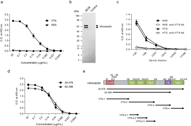
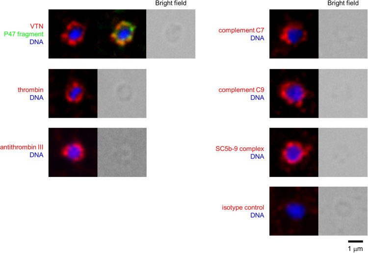
The malaria parasite Plasmodium falciparum proliferates in the blood stream where the host immune system is most active. To escape from host immunity, P. falciparum has developed a number of evasion mechanisms. Serine repeat antigen 5 (SERA5) is a blood stage antigen highly expressed at late trophozoite and schizont stages. The P47 N-terminal domain of SERA5, the basis of SE36 antigen of the blood stage vaccine candidate under clinical trials, covers the merozoite surface. Exploring the role of the P47 domain, screening of serum proteins showed that vitronectin (VTN) directly binds to 20 residues in the C-terminal region of SE36. VTN co-localized with P47 domain in the schizont and merozoite stages. Phagocytosis assay using THP-1 cells demonstrated that VTN bound to SE36 prevented engulfment of SE36-beads. In addition, several serum proteins localized on the merozoite surface, suggesting that host proteins camouflage merozoites against host immunity via binding to VTN.


Conflict of interest statement

The authors declare no competing interests.

Figures

Figure 1
Figure 1
Binding assay of vitronectin (VTN) to SE36. (a) Reactivity of the purified VTN and HSA against SE36. SE36 was adsorbed to microtiter plate and VTN or human serum albumin (HSA) was added at the indicated concentrations. (b) Western blotting of elutes from SE36-immobilized and control columns. The anti-VTN pAb detected two forms (V75 and V65; upper band and lower band, respectively) of VTN. Each elute was run in an SDS-PAGE gel and probed with anti-VTN pAb (diluted 1:2000). (c) Reactivity of VTN in naïve human serum (NHS) and Ugandan high anti-SE36 IgG-titer serum (HTS) against SE36. SE36 was adsorbed to microtiter plate and NHS or HTS was added at the indicated dilutions (closed symbols). Open symbols show reactions without anti-VTN Ab as negative control. (d) Reactivity of commercially available VTN recombinants (see also Fig. 1e) against SE36. SE36 was adsorbed to microtiter plate and VTN recombinants were added at the indicated concentrations. (e) Schematic representation of VTN. “SmB”, “Hp”, and “Hb” indicate Somatomedin-B motif, Hemopexin domain, and Heparin-binding region, respectively. The number 398 denotes the endogenous cleavage site. The designations “20–478” and “62–398” indicate commercially available VTN recombinants. VTN-1 to -4, as well as VTN-2-1 to -2-3 denote truncated recombinant forms. Results in (ac, and d) are expressed as means ± SD from three independent experiments. In (c), statistical analysis between NHS and HTS was performed using a Mann-Whitney U test. No significant differences were found.
Figure 2
Figure 2
Mapping of binding site of VTN in SE36. (a) Schematic representation of SE36. The “8” and “S” indicate octamer repeat and serine rich region, respectively. “RGD” indicates RGD motif. The number 178 denotes the position of polyserine sequence present in PfSERA5 but deleted in SE36. SE36-1 to -4 denote truncated recombinant forms. P1 to P15 are synthetic peptides. (b) Reactivity of VTN in NHS and HTS against each truncated SE36 recombinant. Each recombinant was adsorbed to microtiter plate and NHS and HTS were added at 1:2000 dilution. (c) Reactivity of the purified VTN against each truncated SE36 recombinant. Each recombinant was adsorbed to microtiter plate and the purified VTN was added at 2 μg/mL concentration. (d) Reactivity of VTN in NHS and HTS against each synthetic peptide. Each synthetic peptide was adsorbed to microtiter plate (0.3 μM) and NHS and HTS were added at 1:2000 dilution. (e) Reactivity of the purified VTN against each synthetic peptide. Each synthetic peptide was adsorbed to microtiter plate and the purified VTN was added at 2 μg/mL concentration. Results in (be) are expressed as means ± standard deviation (SD) from three independent experiments. In (b and d), statistical analyses between NHS and HTS were performed using a Mann-Whitney U test. No significant differences were found.
Figure 3
Figure 3
Localization of VTN and P47 domain/SERA5. (a) (i) Representative immunofluorescence assay (IFA) images of merozoites and schizont. (ii) Upper panels, Representative IFA images of schizont. VTN was probed with an isotype control mAb instead of anti-VTN mAb. Lower panels, Representative IFA images of schizont cultured in AlbuMAX-I medium. Target proteins were probed with anti-VTN mAb (green), anti-SE36 rabbit serum (red), and DAPI nuclear stain (blue). Scale bar, 5 μm. (b) Representative IFA images of merozoite and schizont stages under deconvolution microscopy. Probes used were the same as in panel a. Upper panels show the localization of P47 domain/SERA5 during merozoite and schizont stages. Lower panels show the intensity of green and red signals at the section of the gray line in the upper panel. Scale bar, 1 μm. (c) Western blotting of NHS, red blood cell (RBC) lysate, and iRBC lysate. The blots were probed with anti-SE36 mouse serum (diluted 1:1000), anti-VTN pAb (diluted 1:2000), anti-albumin pAb (diluted 1:2000), and anti-band 3 mAb (diluted 1:2000). Band 3 is RBC-specific protein and used as loading control. (d) Representative IFA images of early trophozoite and trophozoite. Target proteins were probed with anti-VTN mAb (green), anti-SE36 rabbit serum (red), and DAPI nuclear stain (blue). Scale bar, 5 μm.
Figure 4
Figure 4
Characterization of VTN binding to SE36. (a) Biophysical interaction analysis of the binding of VTN to SE36 using surface plasmon resonance (SPR). The raw data (color lines) were fitted to 1:1 binding and bivalent analyte model (black lines) using the Biacore T200 Evaluation software. The fit parameters from these models are summarized in (b). These experiments were performed in triplicate, and representative data are shown. (b) Kinetic and equilibrium constants for the binding of VTN to SE36. (c) Schematic representation of two schemes in bivalent binding model. (i) VTN contains two binding sites. (ii) VTN contains a single binding site and forms a dimer. (d) (i) Purity of VTN-1 to -4 recombinants confirmed by sodium dodecyl sulphate-polyacrylamide gel electrophoresis (SDS-PAGE) and Coomassie Brilliant Blue (CBB) staining. Two micrograms of each recombinant was run in a gel. Arrows indicate target proteins. Since estimated molecular weights were 18, 21, 14, and 12 kDa in VTN-1 to -4, respectively, it was presumed that these recombinants were glycosylated in Pichia pastoris. (ii) Reactivity of SE36 against each truncated VTN recombinant. Each recombinant was adsorbed to microtiter plate (1 μg/mL) and SE36 was added at the indicated concentrations. (e) (i) Purity of VTN-2-1 to -2-3 recombinants confirmed by SDS-PAGE and CBB staining. Two micrograms of each of recombinant was run in a gel. Arrows indicate target proteins. Since estimated molecular weights were 8 kDa in VTN-2-1 to -2-3, it was, likewise, presumed that these recombinants were glycosylated in P. pastoris. All recombinant proteins of VTN (d and e) were detected by anti-His tag antibody in western blotting and was purified by His GraviTrap. (ii) Reactivity of SE36 against each truncated VTN recombinant. Each recombinant was adsorbed to microtiter plate (1 μg/mL) and SE36 was added at the indicated concentrations.
Figure 5
Figure 5
Phagocytosis assay by THP-1 cells. (a) (i) Western blotting of bound protein(s) on latex beads after solubilization. Lane 1: SE36-beads, lane 2: SE36-beads with VTN, lane 3: latex-beads alone, lane 4: beads with VTN. SE36 and VTN bound to the beads were detected by anti-SE36 mouse serum (diluted 1:1000) and anti-VTN pAb (diluted 1:2000), respectively. Arrows indicate the target proteins. (ii) Representative flow cytometry (FCM) histogram of the antibody-independent engulfment of SE36-beads with or without VTN by THP-1 cells. (iii) Giemsa staining of engulfed SE36-beads (left panel) and non-treated beads (right panel) by THP-1 cells. Arrows indicate the engulfed SE36-beads. Scale bar, 10 μm. (b) FCM histogram of the engulfment by THP-1 cells of SE36-beads with or without VTN and/or purified IgG from NHS or HTS. (c) FCM histogram of the engulfment by THP-1 cells of SE36-beads treated with normal serum or VTN-depleted serum (VTN-depl). (d) FCM histogram of the engulfment by THP-1 cells of SE36-beads with or without VTN-depl, with or without purified VTN or with normal serum.
Figure 6
Figure 6
Localization of serum proteins that bind to VTN. IFA images showing the localization of VTN, thrombin, antithrombin III, complement C7, complement C9, SC5b-9 complex (red), P47 fragment (green), and DNA (blue) on the merozoite. Scale bar, 1 μm.

Similar articles

Cited by

References

    1. Bhatt S, et al. The effect of malaria control on Plasmodium falciparum in Africa between 2000 and 2015. Nature. 2015;526:207–211. doi: 10.1038/nature15535. - DOI - PMC - PubMed
    1. Richards JS, Beeson JG. The future for blood-stage vaccines against malaria. Immunol. Cell Biol. 2009;87:377–390. doi: 10.1038/icb.2009.27. - DOI - PubMed
    1. Chan JA, Fowkes FJ, Beeson JG. Surface antigens of Plasmodium falciparum-infected erythrocytes as immune targets and malaria vaccine candidates. Cell Mol. Life Sci. 2014;71:3633–3657. doi: 10.1007/s00018-014-1614-3. - DOI - PMC - PubMed
    1. Pérez Arellano JL, Espinoza EY, Sánchez MM, Muro A. Evasion mechanisms of parasites. Revista Ibérica de Parasitología. 2001;61:4–16.
    1. Ibáñez-Escribano A, et al. Sequestration of host-CD59 as potential immune evasion strategy of Trichomonas vaginalis. Acta Trop. 2015;149:1–7. doi: 10.1016/j.actatropica.2015.05.003. - DOI - PubMed

Publication types

MeSH terms