. 2018 Apr 1;9(4):436.
doi: 10.1038/s41419-018-0402-7.

Nuclear receptor binding protein 1 correlates with better prognosis and induces caspase-dependent intrinsic apoptosis through the JNK signalling pathway in colorectal cancer

Affiliations

Nuclear receptor binding protein 1 correlates with better prognosis and induces caspase-dependent intrinsic apoptosis through the JNK signalling pathway in colorectal cancer

Yi Liao et al. Cell Death Dis. .

Abstract

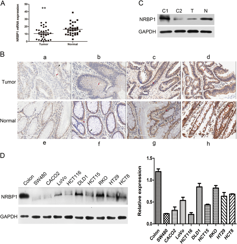
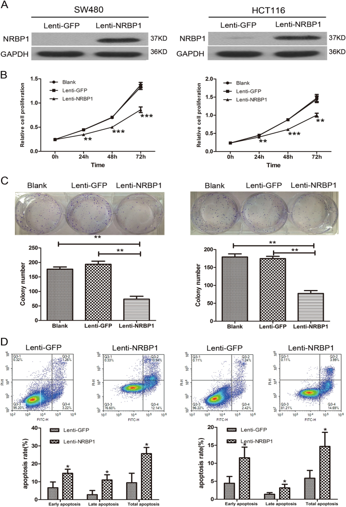
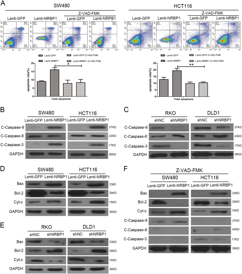
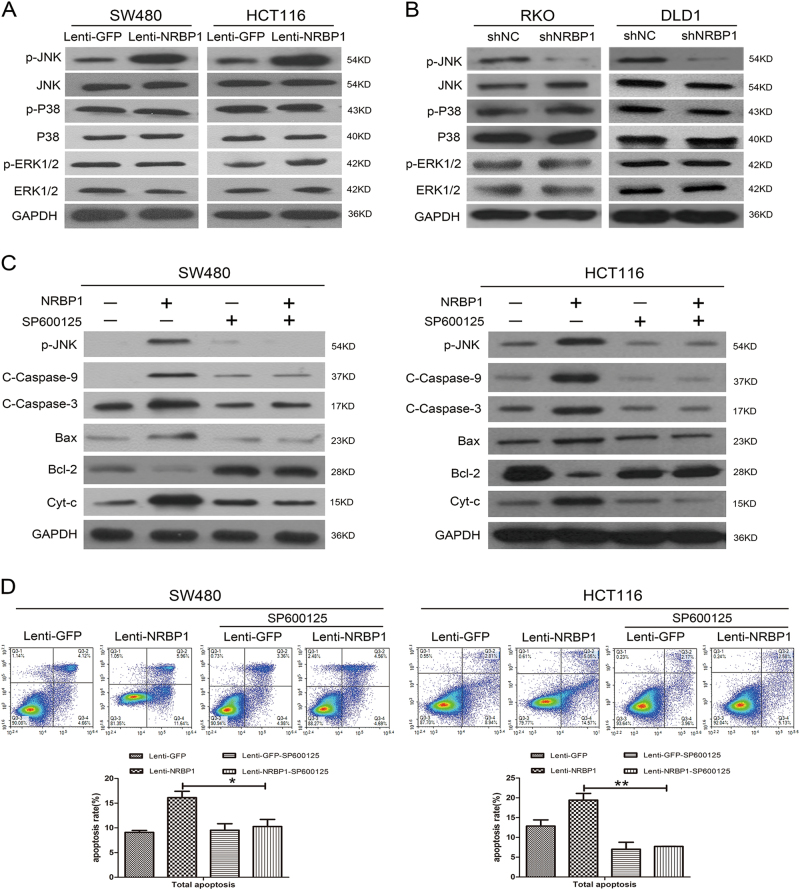
Nuclear receptor binding protein 1 (NRBP1) is a ubiquitously expressed and highly conserved pseudokinase that has important roles in cellular homoeostasis. Despite recent advances in understanding the biology of NRBP1, the role of NRBP1 and its underlying mechanism in colorectal cancer (CRC) have not been fully elucidated. In the present study, we observed that NRBP1 expression levels were significantly reduced in CRC tissues compared with corresponding adjacent normal tissues, and high NRBP1 expression correlated with better prognosis in CRC. Overexpression of NRBP1 inhibited CRC cell proliferation and promoted apoptosis in vitro and in vivo. In contrast, knockdown of NRBP1 expression increased cell proliferation and decreased the percentage of apoptotic cells. Moreover, overexpression of NRBP1 activated caspase-dependent intrinsic apoptosis. In addition, we further discovered that NRBP1 regulated the apoptotic pathway through interaction with JNK. Finally, NRBP1 overexpression led to attenuated CRC growth in a xenograft mouse model. Our study illustrates the suppressor role of NRBP1 in CRC and provides a potential therapeutic target.


Conflict of interest statement

The authors declare that they have no conflict of interest.

Figures

Fig. 1
Fig. 1. Expression of NRBP1 is downregulated in CRC.
a qRT-PCR analysis of the expression levels of NRBP1 in 30 pairs of fresh-frozen primary CRC tissues and their matched normal tissues adjacent to cancer tissues. Three experiments were done. NRBP1 mRNA expression was normalised to β-actin mRNA, which used as the internal reference. The data are expressed as the mean ± standard deviation. (**P < 0.01). b IHC staining to detect NRBP1. a Weak staining of NRBP1 in CRC tissues. bd More intense staining of NRBP1 in CRC tissues. e Weak staining of NRBP1 in normal colorectal tissues. fh More intense staining of NRBP1 in normal colorectal tissues. Red arrow indicates cytoplasm staining. Black arrow indicates nucleus staining. c The expression levels of NRBP1 protein were analysed by western blot in CRC tissue and matched normal colorectal tissue. GAPDH was used as loading control. N normal tissue, T tumour tissue, C1 protein from stomach tissue was used as positive control, C2 protein from thymus tissue was used as negative control. d Western blot was used to investigate NRBP1 protein expression in nine CRC cell lines and normal colorectal tissue
Fig. 2
Fig. 2. High NRBP1 expression correlate with better prognosis in CRC.
Kaplan–Meier curves displaying a overall survival and b disease-free survival in patients with primary CRC based on NRBP1 expression status (high or low expression). High expression patients had better survival than low expression patients. P values were defined by log-rank test
Fig. 3
Fig. 3. Effects of ectopic expression of NRBP1 on proliferation and apoptosis of SW480 and HCT116 cells.
a Increase levels of NRBP1 in SW480 and HCT116 cells transduced with lenti-NRBP1 was confirmed by western blot. b The short time effect of ectopic NRBP1 on the tumour cell proliferation was evaluated by CCK-8 at 0, 24, 48 and 72 h. c Representative pictures (top) of colony formation assay for crystal violet-stained cells and quantified analysis (bottom). Values represent three independent experiments. d Cell apoptosis was measured by flow cytometry analysis following Annexin V and PI staining. Experiment was repeated three times in triplicate. Images show a representative result (top). Q3-1 necrosis cells. Q3-2 late apoptosis cells. Q3-3 normal cells. Q3-4 early apoptosis cells. Graphs (bottom) show quantitative analysis of early apoptosis, late apoptosis and total apoptosis (*P < 0.05, **P < 0.01, ***P < 0.001)
Fig. 4
Fig. 4. Effects of knockdown of NRBP1 on proliferation and apoptosis of RKO and DLD1 cells.
a Decreased levels of NRBP1 in RKO and DLD1 cells transduced with sh2 and sh3 was confirmed by western blot. b The short time effect of silencing NRBP1 on the tumour cell proliferation was evaluated by CCK-8 at 0, 24, 48 and 72 h. c Representative pictures (top) of colony formation assay for crystal violet-stained cells and quantified analysis (bottom). Values represent three independent experiments. d Cell apoptosis was measured by flow cytometry analysis following Annexin V and PI staining. Experiment was repeated three times in triplicate. Images shows a representative result (top). Q3-1 necrosis cells. Q3-2 late apoptosis cells. Q3-3 normal cells. Q3-4 early apoptosis cells. Graphs (bottom) show quantitative analysis of early apoptosis, late apoptosis and total apoptosis (*P < 0.05, **P < 0.01, ***P < 0.001)
Fig. 5
Fig. 5. NRBP1 regulates caspase-dependent intrinsic apoptosis in CRC cells.
a SW480 and HCT116 cells were treated with Z-VAD-FMK for 1 h, a pan-caspase inhibitor. Twenty-four hours later, apoptotic ratios were analysed by double staining the cells with Annexin V and PI using flow cytometry. Representative images from triplicate experiments are shown (top). Q3-1 necrosis cells. Q3-2 late apoptosis cells. Q3-3 normal cells. Q3-4 early apoptosis cells. Graphs (bottom) show quantitative analysis of total apoptosis. b Western blot was performed to investigate the expression levels of caspase-related proteins (caspase-9, caspase-8, caspase-3) in SW480 and HCT116 cells transduced with lenti-NRBP1 (lenti-GFP was used as a negative control). GAPDH was used as loading control. c Western blot was performed to investigate the expression levels of caspase-related proteins (caspase-9, caspase-8, caspase-3) in RKO and DLD1 cells transduced with shNRBP1. GAPDH was used as loading control. d Bax, Bcl-2, cytosolic cytochrome c and GAPDH (loading control) protein levels were detected by western blot in SW480 and HCT116 cells transduced with lenti-NRBP1 (lenti-GFP was used as a negative control). e Bax, Bcl-2, cytosolic cytochrome c and GAPDH (loading control) protein levels were detected by western blot in RKO and DLD1 cells transduced with shNRBP1. f The effects of Z-VAD-FMK on caspase-related protein in SW480 and HCT116 cells transduced with lenti-NRBP1 or lenti-GFPS were evaluated by western blot analysis (*P < 0.05, **P < 0.01)
Fig. 6
Fig. 6. NRBP1 promotes apoptosis through the activation of JNK in CRC cells.
a The effects of NRBP1 overexpression on targets in the JNK, P38, ERK1/2 signalling pathways were assessed by western blot in SW480 and HCT116 cells. GAPDH was used as loading control. b The effects of NRBP1 silencing on targets in the JNK, P38, ERK1/2 signalling pathways were assessed by western blot in RKO and DLD1 cells. GAPDH was used as loading control. c SW480 and HCT116 cells were treated with SP600125 for 30 min. Twenty-four hours later, caspase-related proteins were evaluated by western blot. c Apoptotic ratios were analysed by double staining the cells with Annexin V and PI using flow cytometry. Representative images from triplicate experiments are shown (top). Q3-1 necrosis cells. Q3-2 late apoptosis cells. Q3-3 normal cells. Q3-4 early apoptosis cells. Graphs (bottom) show quantitative analysis of total apoptosis (*P < 0.05, **P < 0.01)
Fig. 7
Fig. 7. Inhibition of tumour growth by NRBP1 in vivo, and association of NRBP1 and apoptosis-related proteins in CRC tissue specimens and cell lines.
a Female nude were subcutaneously injected with SW480-NRBP1 and SW480-GFP cells. Tumours were measured using digital calipers every 5 days. Tumour volume was calculated and recorded. The mean volumes in mice injected with SW480-NRBP1 cells were significantly smaller than in the controls. b On day 25 after injection, mice were killed and the tumours were removed and weighed. Image shows the photograph of representative xenograft tumours of two different treatment groups. c Expression of PCAN, cleaved caspase-9, cleaved caspase-3 and phosphorylated JNK was determined by IHC staining in tumour xenografts. Cell apoptosis was analysed by TUNEL staining. d Expression of phosphorylated JNK and cleaved caspase-9 was determined by IHC staining in CRC tissues. e The expression of phosphorylated JNK in the nine human CRC cells was assayed by western blot. The expression of GAPDH was used as loading control. (**P < 0.01, ***P < 0.001)

Similar articles

Cited by

References

    1. Torre LA, et al. Global cancer statistics, 2012. CA Cancer J. Clin. 2015;65:87–108. doi: 10.3322/caac.21262. - DOI - PubMed
    1. Chen W, et al. National cancer incidence and mortality in China, 2012. Chin. J. Cancer Res. 2016;28:1–11. doi: 10.21147/j.issn.1000-9604.2016.06.02. - DOI - PMC - PubMed
    1. Zheng K, et al. Epigenetic silencing of miR-490-3p promotes development of an aggressive colorectal cancer phenotype through activation of the Wnt/β-catenin signaling pathway. Cancer Lett. 2016;376:178–187. doi: 10.1016/j.canlet.2016.03.024. - DOI - PubMed
    1. Sun, J., Ding, W., Zhi, J. & Chen, W. MiR-200 suppresses metastases of colorectal cancer through ZEB1. Tumor Biol. 37, 15501–15507 (2016). 10.1007/s13277-015-3822-3. - PubMed
    1. Burgess AW, Faux MC, Layton MJ, Ramsay RG. Wnt signaling and colon tumorigenesis—a view from the periphery. Exp. Cell Res. 2011;317:2748–2758. doi: 10.1016/j.yexcr.2011.08.010. - DOI - PubMed

Publication types

MeSH terms