. 2018 Apr 1;9(4):432.
doi: 10.1038/s41419-018-0409-0.

BPIFB1 (LPLUNC1) inhibits radioresistance in nasopharyngeal carcinoma by inhibiting VTN expression

Affiliations

BPIFB1 (LPLUNC1) inhibits radioresistance in nasopharyngeal carcinoma by inhibiting VTN expression

Fang Wei et al. Cell Death Dis. .

Abstract

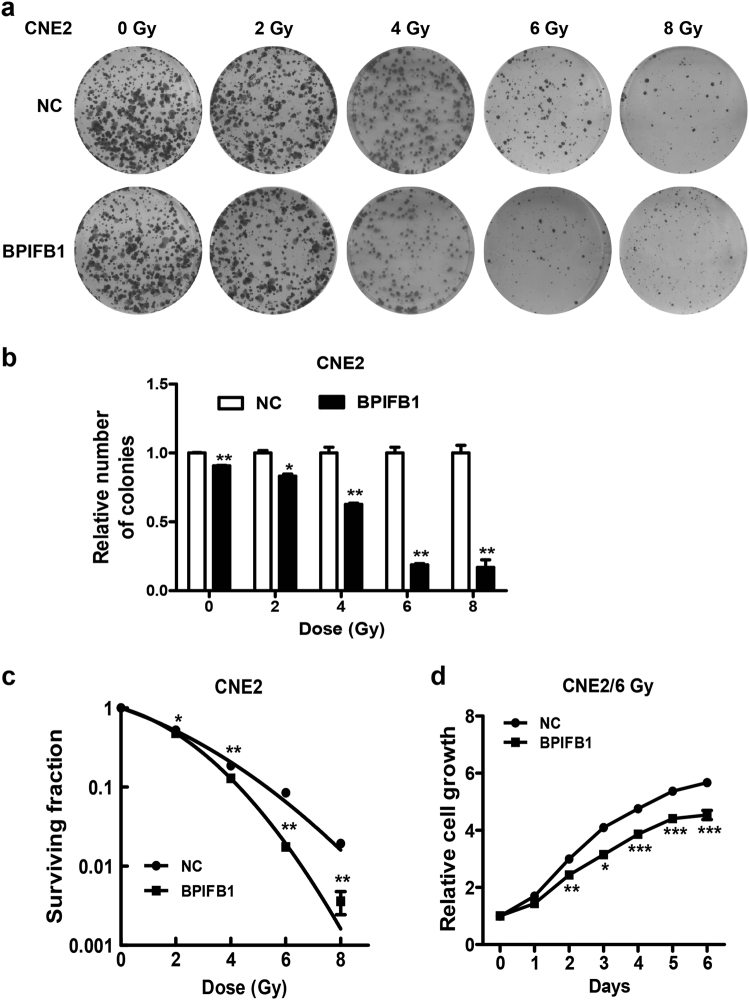
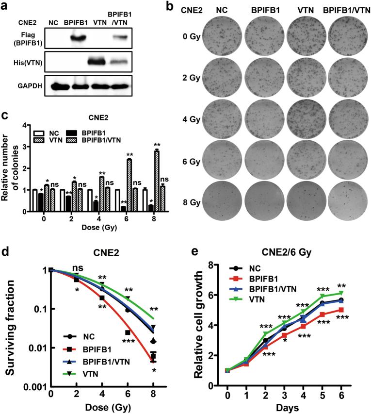
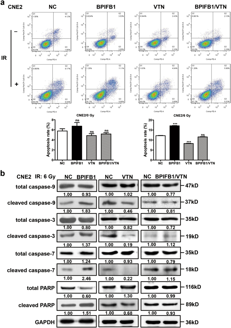
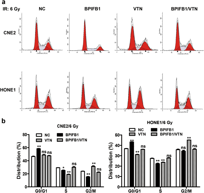
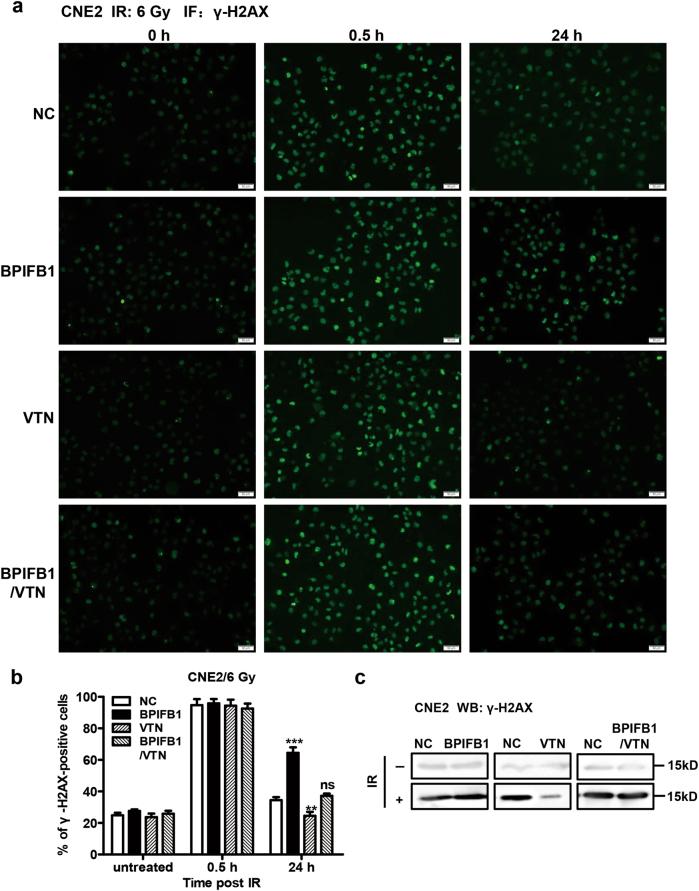
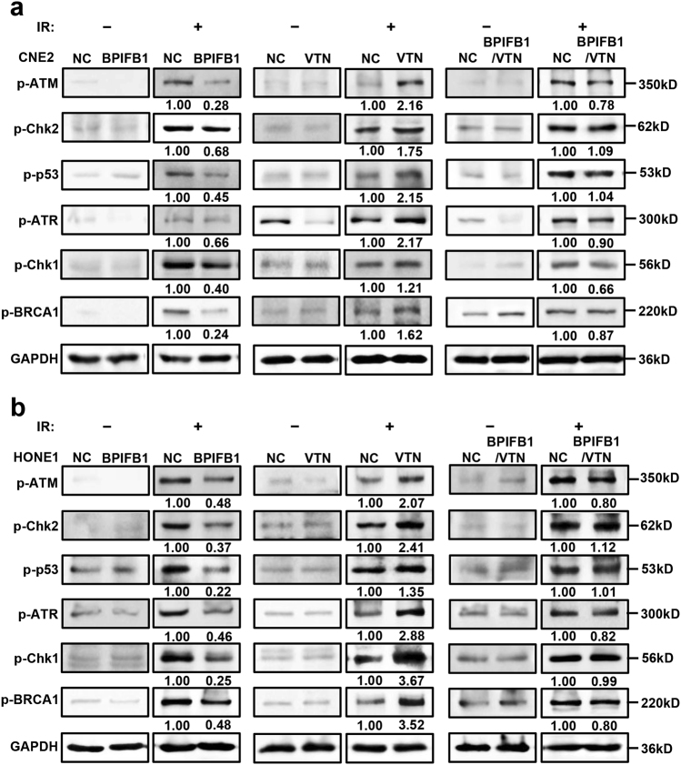
Bactericidal/permeability-increasing-fold-containing family B member 1 (BPIFB1, previously named LPLUNC1) is highly expressed in the nasopharynx and significantly downregulated in nasopharyngeal carcinoma (NPC). Low expression is also associated with poor prognosis in patients with NPC. Radiotherapy is a routine treatment for NPC; however, radioresistance is a major cause of treatment failure. Thus, we aimed to investigate the role of BPIFB1 in the radioresponse of NPC. Colony formation and cell survival results showed that BPIFB1 sensitized NPC cells to ionizing radiation. VTN, a previously identified BPIFB1-binding protein, was shown to induce cell proliferation and survival, G2/M phase arrest, DNA repair, activation of the ATM-Chk2 and ATR-Chk1 pathways, and anti-apoptotic effects after exposure to radiation, facilitating NPC cell radioresistance. However, BPIFB1 inhibited this VTN-mediated radioresistance, ultimately improving NPC radiosensitivity. In conclusion, this study is the first to demonstrate the functions of BPIFB1 and VTN in the NPC radioresponse. Our findings indicated that promoting BPIFB1 expression and targeting VTN might represent new therapeutic strategies for NPC.


Conflict of interest statement

The authors declare that they have no conflict of interest.

Figures

Fig. 1
Fig. 1. BPIFB1 re-expression sensitized CNE2 nasopharyngeal carcinoma (NPC) cells to ionizing radiation (IR).
a Effects of BPIFB1 on clone formation ability of CNE2 cells after irradiation with a dose of 0–8 Gy. b Numbers of surviving foci are presented as bar graphs representing means ± SD. c Effects of BPIFB1 on survival rate of CNE2 cells after radiotherapy. Surviving fractions were calculated as described based on the data from experiments depicted in b. d CCK8 assays were used to investigate the role of BPIFB1 in CNE2 cell proliferation before and after irradiation. *P < 0.05; **P < 0.01; ***P < 0.001
Fig. 2
Fig. 2. BPIFB1 regulated the CNE2 nasopharyngeal carcinoma cell radioresponse by interacting with VTN.
a BPIFB1 and VTN expression were confirmed by western blotting in CNE2 cells transfected or co-transfected with BPIFB1-Flag and VTN-His vectors, using anti-Flag and anti-His primary antibodies. b Clone formation assay showing the response of CNE2-NC (negative control), CNE2-BPIFB1, CNE2-VTN, and CNE2-BPIFB1/VTN cells to 0–8 Gy radiotherapy. c The numbers of surviving foci in the four groups are presented as bar graphs representing means ± SD. d Effects of BPIFB1 and VTN on survival of CNE2 cells after radiotherapy. Surviving fractions were calculated as described based on the data from experiments in c. e CCK8 assays were used to investigate the role of BPIFB1 and VTN in CNE2 cell proliferation after irradiation. *P< 0.05; **P< 0.01; ***P< 0.001; NS, no significance
Fig. 3
Fig. 3. Overexpression of BPIFB1 inhibited VTN-induced anti-apoptotic effects in CNE2 nasopharyngeal carcinoma cells after irradiation.
a (Top) BPIFB1-overexpressing cells are sensitive to IR-induced cell death. The four groups of CNE2 cells were treated with or without 6 Gy of ionizing radiation and cells were stained with annexin V to measure the percentage of apoptotic cells. (Below) The percentage of apoptotic cells is presented as bar graphs representing means ± SD. ***P< 0.001; NS, no significance. b Expression of typical apoptosis markers, including cleaved caspase-9, cleaved caspase-3, cleaved caspase-7, and cleaved PARP, based on western blotting, comparing the four groups of CNE2 cells transfected or co-transfected with the BPIFB1-Flag and VTN-His vectors. GAPDH was used as an internal control. NC negative control. The numbers below blots represent grayscale values of each blot. The molecular weights of blots are indicated to their right
Fig. 4
Fig. 4. Overexpression of BPIFB1 impaired VTN-induced nasopharyngeal carcinoma cell cycle arrest after irradiation.
Flow cytometry was performed to assess variations in cell cycle distribution. a BPIFB1 overexpression resulted in G0/G1 phase arrest, and VTN overexpression resulted in G2/M phase arrest in CNE2 and HONE1 cells after irradiation. The four groups of cells (overexpressing BPIFB1, VTN, both, and the control group) were incubated for 24 h after treatment with 6 Gy of radiation. b Histogram of cell cycle distribution of CNE2 and HONE1 cells. Data represent means ± SD from of at least three independent experiments; *P < 0.05; **P < 0.01; NS, no significance
Fig. 5
Fig. 5. BPIFB1 repressed VTN-induced DNA repair in CNE2 nasopharyngeal carcinoma cells after irradiation.
The levels of γ-H2AX at different times after 6 Gy irradiation were detected by immunofluorescence in CNE2 cells transfected or co-transfected with BPIFB1-Flag and VTN-His vectors. Cells displaying 10 or more foci were counted as positive. a Representative images of γ-H2AX foci in CNE2-NC (negative control), CNE2-BPIFB1, CNE2-VTN, and CNE2-BPIFB1/VTN cells are shown. **P< 0.01; ***P< 0.001; NS, no significance. Scale bar = 50 μm. b Histogram of the percentage of γ-H2AX foci in the four groups of CNE2 cells. c Detection of γ-H2AX protein levels in the four groups of CNE2 cells treated with or without 6 Gy of IR. The molecular weights of blots are indicated to their right
Fig. 6
Fig. 6. Effects of BPIFB1 and VTN on ionizing radiation (IR)-induced activation of the ATM/Chk2 and ATR/Chk1 pathways in nasopharyngeal carcinoma (NPC) cells.
Expression of ATM/Chk2 and ATR/Chk1 pathway-associated proteins. Protein levels of phospho (p)-ATM, p-Chk2, p-p53, p-ATR, p-Chk1, and p-BRCA1 were detected by western blotting in a CNE2 cells and b HONE1 cells transfected or co-transfected with BPIFB1-Flag and VTN-His vectors. NC, negative control. The numbers below blots represent grayscale values of each blot. The molecular weights of blots are indicated to their right

Similar articles

Cited by

References

    1. Wei F, et al. Trend analysis of cancer incidence and mortality in China. Sci. China Life Sci. 2017;60:1–4. - PubMed
    1. He B, et al. Epstein–Barr virus-encoded miR-BART6-3p inhibits cancer cell metastasis and invasion by targeting long non-coding RNA LOC553103. Cell Death Dis. 2016;7:e2353. doi: 10.1038/cddis.2016.253. - DOI - PMC - PubMed
    1. Lo KW, To KF, Huang DP. Focus on nasopharyngeal carcinoma. Cancer Cell. 2004;5:423–428. doi: 10.1016/S1535-6108(04)00119-9. - DOI - PubMed
    1. Chua ML, et al. Nasopharyngeal carcinoma. Lancet. 2016;387:1012–1024. doi: 10.1016/S0140-6736(15)00055-0. - DOI - PubMed
    1. Razak AR, et al. Nasopharyngeal carcinoma: the next challenges. Eur. J. Cancer. 2010;46:1967–1978. doi: 10.1016/j.ejca.2010.04.004. - DOI - PubMed

Publication types

MeSH terms