. 2018 Mar 21;7(1):41.
doi: 10.1038/s41426-018-0031-3.

Human transbodies that interfere with the functions of Ebola virus VP35 protein in genome replication and transcription and innate immune antagonism

Affiliations

Human transbodies that interfere with the functions of Ebola virus VP35 protein in genome replication and transcription and innate immune antagonism

Watee Seesuay et al. Emerg Microbes Infect. .

Abstract

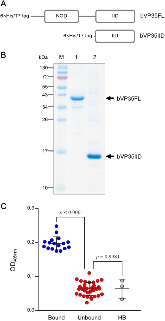
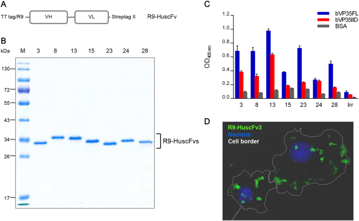
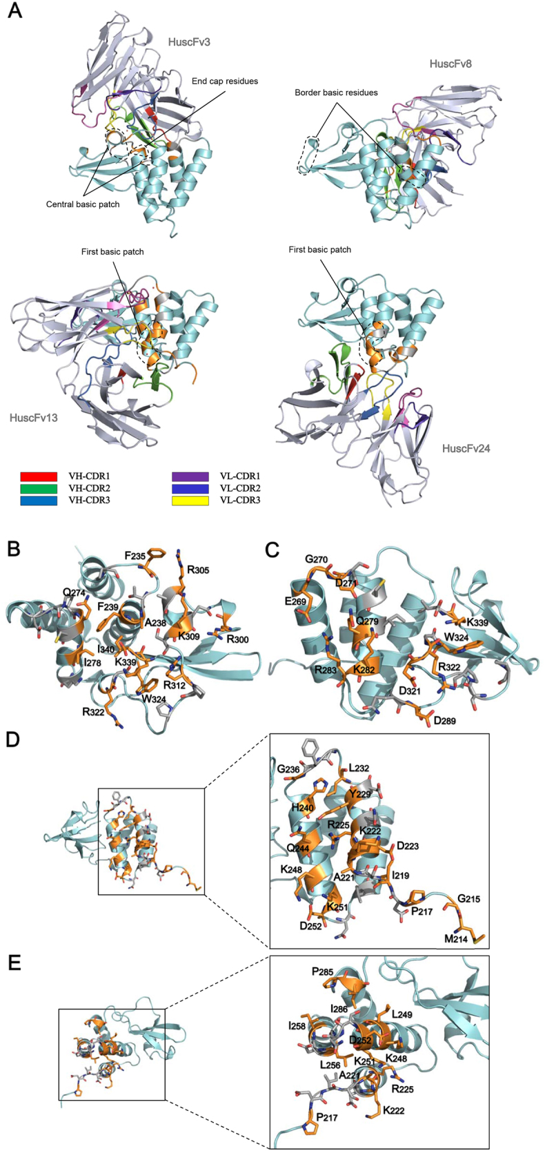
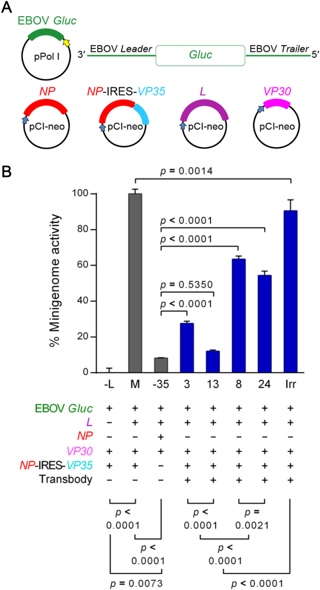
Small molecular inhibitors and passive immunization against Ebola virus disease (EVD) have been tested in animal models, including rodents and non-human primates, as well as in clinical trials. Nevertheless, there is currently no Food and Drug Administration (FDA)-approved therapy, and alternative strategies must be pursued. The aim of this study was to produce cell-penetrable human single-chain antibodies (transbodies) that are able to interfere with the activities of interferon inhibitory domain (IID) of the VP35 protein, a multifunctional virulence factor of Ebola virus (EBOV). We speculated that effective VP35-IID-specific transbodies could inspire further studies to identify an alternative to conventional antibody therapies. Phage display technology was used to generate Escherichia coli-derived human single-chain antibodies (HuscFvs) that bind to IID. HuscFvs were linked to nona-arginine (R9) to make them cell penetrable. Transbodies of transformed E. coli clones 13 and 3, which were predicted to interact with first basic patch residues (R9-HuscFv13), central basic patch, and end-cap residues (R9-HuscFv3), effectively inhibited EBOV minigenome activity. Transbodies of E. coli clones 3 and 8 antagonized VP35-mediated interferon suppression in VP35-transduced cells. We postulate that these transbodies formed an interface contact with the IID central basic patch, end-cap, and/or residues that are important for IID multimeric formation for dsRNA binding. These transbodies should be evaluated further in vitro using authentic EBOV and in vivo in animal models of EVD before their therapeutic/prophylactic effectiveness is clinically evaluated.


Conflict of interest statement

The authors declare that they have no conflict of interest.

Figures

Fig. 1
Fig. 1. Productions of recombinant VP35 proteins and selection of VP35-bound HuscFvs.
a Schematic representations of constructs of bacterially produced recombinant full-length EBOV VP35 (bVP35FL) and the C-terminal interferon inhibitory domain of VP35 (bVP35IID). b Recombinant bVP35FL and bVP35IID proteins purified from transformed E. coli clones. M, pre-stained protein ladder; lane 1, purified bVP35FL; and, lane 2, purified bVP35IID. Numbers at the left represent the protein molecular masses in kDa. c bVP358FL-bound HuscFv clones, as determined by indirect ELISA using purified bVP35FL as antigen. The bound group was selected from the OD405nm signal above mean + 3SD of the background binding control (lysate of original E. coli HB2151; HB). Statistical significance was determined using one-way ANOVA and Tukey’s post hoc test. Supplementary Figure S1 provides details of the binding of individual clones to bVP35FL (test antigen) and BSA (control antigen)
Fig. 2
Fig. 2. Antigen binding and cell entry ability of purified R9-HuscFvs.
a Schematic representation of the construct for preparing cell-penetrable HuscFvs (R9-HuscFvs). b SDS-PAGE and CBB-stained R9-HuscFvs purified and refolded from transformed E. coli clones 3, 8, 13, 15, 23, 24, and 28. R9-HuscFvs had a molecular mass of ~34 kDa under reducing condition. c Binding activities of R9-HuscFvs to bVP35FL and bVP35IID compared to BSA (control antigen), as demonstrated by indirect ELISA. Positive binding to the tested antigens yielded an OD405nm signal three times higher than to that of the control antigen. Supplementary Figure S2 shows the EC50 value derived from selected bVP35IID-bound R9-HuscFvs. d Intracellular localization of R9-HuscFv was revealed by confocal immunofluorescence microscopy. HepG2 cells were incubated with R9-HuscFv3 (representative of the R9-HuscFvs) for 3 h, and then the cells were fixed, permeabilized, and stained. Cell border, white line; R9-HuscFv, green; nuclei, blue
Fig. 3
Fig. 3. Computerized-generated structures of VP35-IID-HuscFv complexes and presumptive VP35-IID epitopes.
a Overall structures of EBOV VP35-IID (PDB ID: 3FKE) (cyan) after complexing with HuscFvs (blue white) of E. coli clones 3, 8, 13, and 24 derived from molecular docking. Computer-generated animated images of VP35-IID that formed direct interface contact with antibodies are colored in orange, while the interfaces that fell within 5 Å thresholds of the van der Waals radii of the HuscFvs are colored in gray. b–e Contact interfaces between VP35-IID and HuscFv3, HuscFv8, HuscFv13, and HuscFv24. Residues of the IID that make contact with their respective HuscFvs are shown as sticks. For details, see Supplementary Table S1
Fig. 4
Fig. 4. Interactions of the transbodies with VP35 and their biocompatibility.
a Schematic representation of the VP35 construct for production in mammalian cells (mVP35). b mVP35 produced from COS-7 cells (∼40 kDa; arrow). COS-7 cells were transiently transfected with the VP35 construct. At 48-h post-transfection, the cells were lysed, and the presence of mVP35 in the clarified lysate was determined by Western blotting using anti-Flag antibody. The lysate of cells transfected with empty vector was used as a negative control. c Diagram of co-immunoprecipitation and ELISA for detecting the interaction between mVP35 and R9-HuscFvs. COS-7 cell lysate containing mVP35 was mixed with R9-HuscFvs, and individual mixtures were added to wells containing immobilized anti-Flag antibody. Captured complexes were detected using Strep-Tactin®-HRP conjugate and HRP substrate. d Upper, the OD405nm of ELISA for demonstrating the interaction between mVP35 and R9-HuscFvs compared with mVP35 mixed with irrelevant R9-HuscFv (Irr) and the background binding control (Ctrl; mVP35 without R9-HuscFv). Lower, the input reactants of co-immunoprecipitation revealed by Western blotting are shown. e The biocompatibility of R9-HuscFvs with human hepatic cells shown as the percent viability of HepG2 cells after incubation with R9-HuscFvs for 24 h. The protease activity of dead cells was measured after addition of luminogenic substrate to wells containing treated cells, and the percent cell viability was calculated. Statistical significance was determined using one-way ANOVA and Tukey’s post hoc test
Fig. 5
Fig. 5. Transbody-mediated inhibition of EBOV minigenome activity.
a Schematic diagrams of the plasmids used in the RNA polymerase I-driven EBOV minigenome system. EBOV-like reporter (Gluc) RNA was transcribed under the regulation of human Pol-I promoter and the Sal box transcription termination element. The viral protein expression cassettes were under the regulation of CMV I.E. enhancer/promoter and the SV40 late poly(A) signal element. b Percent EBOV minigenome activity of COS-7 cells after treatment with R9-HuscFvs. COS-7 cells transfected with EBOV minigenome were treated with R9-HuscFv3, R9-HuscFv8, R9-HuscFv13, R9-HuscFv24, and irrelevant R9-HuscFv (Irr). Minigenome activity was measured by detecting Gluc bioluminescent intensity. Controls included (M) COS-7 cells co-transfected with the minigenome without antibody treatment, (−L) COS-7 cells co-transfected with EBOV minigenome without L plasmid, and (−VP35) COS-7 cells co-transfected with the minigenome without VP35 plasmid. Differences in percent minigenome activity were compared using one-way ANOVA and Tukey’s post hoc test
Fig. 6
Fig. 6. INF-β gene response in VP35-transduced cells after treatment with transbodies.
a Schematic diagram of the EBOV VP35 expression cassette (pLVX-VP35) for HepG2 cell transduction. b Conceptual diagram illustrating how the transbodies rescued the IFN-β gene response of host cells from VP35 interferon antagonistic activity. After transduction with lentivector carrying EBOV VP35 gene cassette, transcription of the EBOV-VP35 episome was initiated (cyan solid arrows). The ability of VP35 mRNA to activate MDA5 signaling cascade was antagonized after VP35 production. Cytoplasmic VP35 bioactivity could be blocked by the VP35-targeting transbody (gray solid arrow), which resulted in IFNB1 expression (orange solid arrows) by triggering transgene mRNA. As a consequence, the produced IFN-β induced interferon-stimulated genes (e.g., EIF2AK2 (magenta solid arrow)). Dotted black arrows indicate signaling cascades. c, d Fold change of IFNB1 (c) and EIF2AK2 (d) mRNAs in cells after EBOV VP35 transduction for 12 and 36 h compared to untreated control cells. Cells transfected with poly(I:C) served as positive stimulation, and cells transduced with EBOV VP35H240E and empty vector were transduction controls. Significant differences were determined by two-way ANOVA and the Šidák t test. e, f Expression of IFNB1 (e) and EIF2AK2 (f) mRNAs of untreated control HepG2 cells after incubation with transbodies for 12 h in comparison to the control cells in culture medium alone. g, h Fold change of IFNB1 (g) and EIF2AK2 (h) mRNAs in VP35-transduced cells after exposure to transbodies for 12 h compared with VP35-transduced cells. Controls included untreated cells with and without poly(I:C) transfection, VP35-transduced cells with and without poly(I:C) transfection, and VP35-transduced cells treated with irrelevant transbody (Irr). Statistically significant differences were determined using one-way ANOVA and Dunnett’s post hoc test

Similar articles

Cited by

References

    1. Leung DW, Prins KC, Basler CF, Amarasinghe GK. Ebolavirus VP35 is a multifunctional virulence factor. Virulence. 2010;1:526–531. doi: 10.4161/viru.1.6.12984. - DOI - PMC - PubMed
    1. Mühlberger E, Lötfering B, Klenk HD, Becker S. Three of the four nucleocapsid proteins of Marburg virus, NP, VP35, and L, are sufficient to mediate replication and transcription of Marburg virus-specific monocistronic minigenomes. J. Virol. 1998;72:8756–8764. - PMC - PubMed
    1. Noda T, Aoyama K, Sagara H, Kida H, Kawaoka Y. Nucleocapsid-like structures of Ebola virus reconstructed using electron tomography. J. Vet. Med. Sci. 2005;67:325–328. doi: 10.1292/jvms.67.325. - DOI - PubMed
    1. Basler CF, et al. The Ebola virus VP35 protein functions as a type I IFN antagonist. Proc. Natl Acad. Sci. USA. 2000;97:12289–12294. doi: 10.1073/pnas.220398297. - DOI - PMC - PubMed
    1. Basler CF, García-Sastre A. Viruses and the type I interferon antiviral system: induction and evasion. Int. Rev. Immunol. 2002;21:305–337. doi: 10.1080/08830180213277. - DOI - PubMed

MeSH terms

Substances