. 2018 Mar 22;8(1):5074.
doi: 10.1038/s41598-018-23478-0.

Cellular response to persistent foot-and-mouth disease virus infection is linked to specific types of alterations in the host cell transcriptome

Affiliations

Cellular response to persistent foot-and-mouth disease virus infection is linked to specific types of alterations in the host cell transcriptome

Lingling Han et al. Sci Rep. .

Abstract

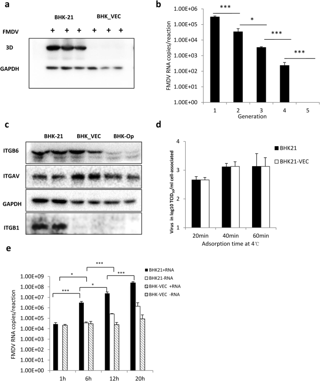
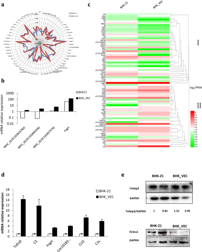
Food-and-mouth disease virus (FMDV) is a highly contagious virus that seriously threatens the development of animal husbandry. Although persistent FMDV infection can dramatically worsen the situation, the mechanisms involved in persistent FMDV infection remain unclear. In the present study, we identified the presence of evolved cells in the persistently FMDV-infected cell line. These cells exhibited resistance to the parent FMDV and re-established persistent infection when infected with FMDV-Op (virus supernatant of persistent infection cell lines), emphasizing the decisive role of evolved host cells in the establishment of persistent FMDV infection. Using RNA-seq, we identified the gene expression profiles of these evolved host cells. In total, 4,686 genes were differentially expressed in evolved cells compared with normal cells, with these genes being involved in metabolic processes, cell cycle, and cellular protein catabolic processes. In addition, 1,229 alternative splicing events, especially skipped exon events, were induced in evolved cells. Moreover, evolved cells exhibited a stronger immune defensive response and weaker MAPK signal response than normal cells. This comprehensive transcriptome analysis of evolved host cells lays the foundation for further investigations of the molecular mechanisms of persistent FMDV infection and screening for genes resistant to FMDV infection.


Conflict of interest statement

The authors declare no competing interests.

Figures

Figure 1
Figure 1
Emergence of FMDV-resistant cells during persistent infection. (a) BHK-21 cells and BHK-VECs were infected with FMDV at 2.5 × 10−4 PFU/cell for 18 h, and whole-cell extracts were analyzed by western blotting using rabbit polyclonal anti-3D antibody and mouse monoclonal anti-GAPDH. (b) BHK-VECs were infected with FMDV at 5 × 10−2 PFU/cell. Infected cells were subcultured, and intracellular viral RNA of each generation was measured using qRT-PCR. (c) Expression of FMDV integrin receptors (ITGB6, ITGAV, ITGB1) in BHK-VEC, BHK-Op, and uninfected BHK-21 cell cultures were detected by western blotting using specific antibodies. (d) Adsorption of FMDV to BHK-21 cells and BHK-VECs. BHK21 cells and BHK-VECs were infected with FMDV at 2.8 PFU/cell and maintained at 0–4 °C. At the indicated time point, supernatant was removed rapidly, and monolayers were washed with cold PBS three times. One mL serum-free MEM was added to the monolayers. Cell suspensions were then subjected to three cycles of freezing and thawing and centrifuged for 5 min at 12,000 × g at 4 °C. Supernatant was used to quantify the bound virus titer by TCID50 assay. (e) Detection of intracellular viral RNA replication in BHK-VECs and BHK-21 cells. BHK21 cells and BHK-VECs were infected with FMDV at 2.5 × 10−4 PFU/cell. At the indicated time points, intracellular RNA was isolated, and intracellular virus RNA (positive-stranded and negative-stranded RNA) was quantified by qRT-PCR. ***p < 0.001, *p < 0.05.
Figure 2
Figure 2
BHK-VECs resisted infection of FMDV-Op. (a) BHK-21 cells and BHK-VECs were infected with FMDV-Op at 2 × 10−4 PFU/cell for 24 h. Whole-cell extracts were analyzed by western blotting using rabbit polyclonal anti-3D-specific or mouse monoclonal anti-GAPDH antibodies. (b) BHK-VECs were infected with FMDV-Op at 2 × 10−4 PFU/cell. Infected cells were subcultured, and the secreted infectious virus in the supernatant of each generation was measured by titration assay. (c) BHK-21 cells were infected with FMDV or FMDV-Op at 2 × 10−4 PFU/cell. Intracellular RNA was isolated for quantification of intracellular virus RNA by qRT-PCR at the indicated time points. ***p < 0.001.
Figure 3
Figure 3
Analysis of DEGs in BHK-VECs. (a) GO enrichment analysis of DEGs in BHK-VECs. We included the top 30 most significant GO categories of DEGs (*p < 0.05). KEGG pathway enrichment analysis was performed for differentially expressed (b) up-regulated and (c) down-regulated genes in BHK-VECs. The graph depicts the most significant 20 enriched pathway entries. (d) Validation of DEGs in BHK-VECs by (i) qRT-PCR or (ii) western blotting. For all qRT-PCR and western blot validations, GAPDH served as the internal reference gene to normalize data.
Figure 4
Figure 4
AS induced in BHK-VECs. (a) Summary of significant differential AS events in BHK-VECs compared with BHK-21 cells. The intron-exon structure involved in each splicing pattern is shown. (be) Verification of AS events by RT-PCR. Visualization of RNA-seq reads was first performed by IGV. Then, RT-PCR was performed using specific primers located upstream and downstream of SE events. The y-axis shows the number of mapped reads, and the red arrows at the bottom show the target area for primer amplification. Hnrnpa2b1, Pde4dip, and Hypk showed increased SEs in BHK-VECs as shown by a larger number of products of 324, 390, and 144 bp. Syt12 showed a decrease in SEs in BHK-VECs as shown by a larger number of products of 227 bp.
Figure 5
Figure 5
Differential expression of immunity-related genes in BHK-VECs. (a) Differential expression of innate immunity-related genes between BHK-21 cells and BHK-VECs were artificially chosen for expression analysis. The x-axis is the periphery of the circle showing the names of genes, and the y-axis is the radius of the circle showing the corresponding FPKM values. The farther away from the center of the circle, the higher the expression of the gene. (b) Comparison of adaptive immunity-related gene expression between BHK-21 cells and BHK-VECs. (c) Hierarchical clustering heatmap of differentially expressed immunity-related genes between BHK-VECs and BHK-21 cells. The color range from green to red corresponds to a low to high abundance of gene expression, respectively. (d,e) Validation of differentially expressed immunity-related genes in BHK-VECs by qRT-PCR or western blotting. GAPDH served as the internal reference gene to normalize data.
Figure 6
Figure 6
Differential expression of MAPK signaling-related genes in BHK-VECs. (a) qRT-PCR validation of DEGs related to MAPK pathways in BHK-VECs. GAPDH served as the internal reference gene to normalize data, and three biologically independent replicates were performed. (b,c) Effect of MAPK/ERK or p38/MAPK inhibition on replication of FMDV in BHK-21 cells. BHK-21 cells were pre-incubated (1 h) with DMSO, (b) 20 or 50 mM U0126, or (c) 20 or 50 mM SB202190 and then infected with FMDV at 2.5 × 10−4 PFU/cell for 24 h in the presence of DMSO, U0126, or SB202190. Protein extracts were examined using western blotting with FMDV 3D-specific or the indicated antibodies; the effectiveness of inhibition was monitored by detecting the phosphorylation of inhibitor-specific target protein (P-MAPK/ERK and pS15-Hsp27). (d) (i) p38δ (MAPK13) was down-regulated in BHK-VECs compared with BHK-21 cells, and (ii) overexpression of MAPK13 genes in BHK-21 cells promoted the replication of FMDV.

Similar articles

Cited by

References

    1. Grubman MJ, Baxt B. Foot-and-mouth disease. Clin Microbiol Rev. 2004;17:465–493. doi: 10.1128/CMR.17.2.465-493.2004. - DOI - PMC - PubMed
    1. Kitching P, et al. Global FMD control–is it an option? Vaccine. 2007;25:5660–5664. doi: 10.1016/j.vaccine.2006.10.052. - DOI - PubMed
    1. Thompson D, et al. Economic costs of the foot and mouth disease outbreak in the United Kingdom in 2001. Revue scientifique et technique. 2002;21:675–687. doi: 10.20506/rst.21.3.1353. - DOI - PubMed
    1. Alexandersen S, Zhang Z, Donaldson AI. Aspects of the persistence of foot-and-mouth disease virus in animals–the carrier problem. Microbes and infection. 2002;4:1099–1110. doi: 10.1016/S1286-4579(02)01634-9. - DOI - PubMed
    1. Moonen P, Schrijver R. Carriers of foot-and-mouth disease virus: a review. The Veterinary quarterly. 2000;22:193–197. doi: 10.1080/01652176.2000.9695056. - DOI - PubMed

Publication types

MeSH terms