. 2018 Mar 22;8(1):5014.
doi: 10.1038/s41598-018-23396-1.

Comparison of the Biological Characteristics of Mesenchymal Stem Cells Derived from the Human Placenta and Umbilical Cord

Affiliations

Comparison of the Biological Characteristics of Mesenchymal Stem Cells Derived from the Human Placenta and Umbilical Cord

Mingjun Wu et al. Sci Rep. .

Abstract

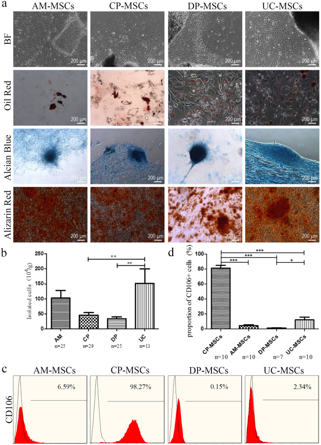
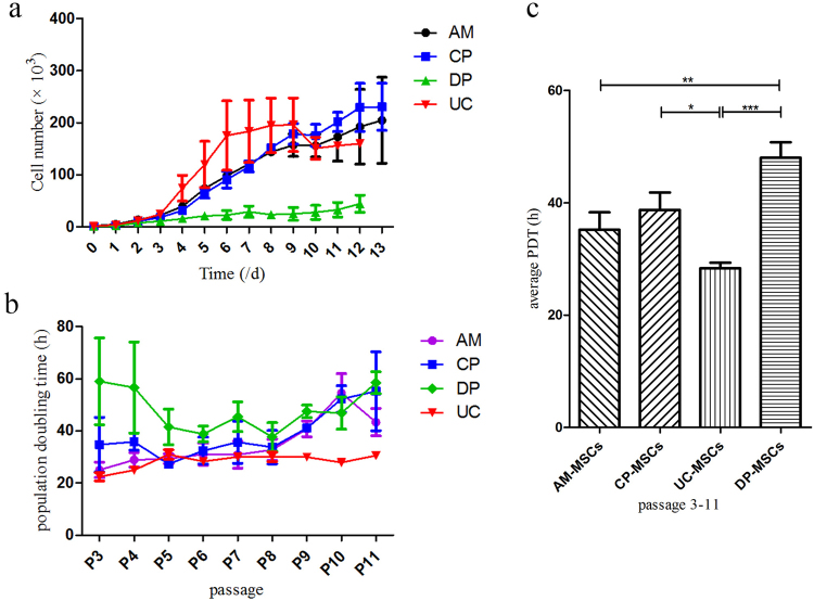
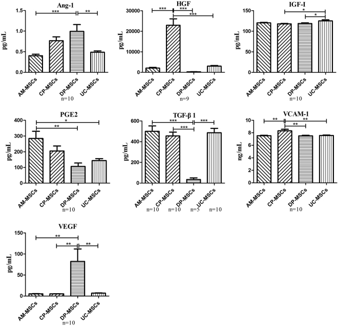
Mesenchymal stem/stromal cells (MSCs) derived from placental tissue show great therapeutic potential and have been used in medical treatment, but the similarity and differences between the MSCs derived from various parts of the placenta remain unclear. In this study, we compared MSCs derived from different perinatal tissues, including the umbilical cord (UC), amniotic membrane (AM), chorionic plate (CP) and decidua parietalis (DP). Using human leukocyte antigen (HLA) typing and karyotype analysis, we found that the first three cell types were derived from the foetus, while the MSCs from the decidua parietalis were derived from the maternal portion of the placental tissue. Our results indicate that both foetal and maternal MSCs share a similar phenotype and multi-lineage differentiation potential, but foetal MSCs show a significantly higher expansion capacity than do maternal MSCs. Furthermore, MSCs from all sources showed significant differences in the levels of several paracrine factors.


Conflict of interest statement

The authors declare no competing interests.

Figures

Figure 1
Figure 1
Characterization and isolation yield of different types of MSCs derived from perinatal tissues. (a) All MSCs exhibited a similar morphology and became positive for oil red O (adipocytic differentiation), alcian blue (chondrocytic differentiation), and alizarin red (osteocytic differentiation). (b) Original raw material, MSC isolation yield. Data are presented as the mean ± SEM (*p < 0.05, **p < 0.005). (c) Flow cytometric analysis of CD106 expression in different MSCs. (d) Statistical result of CD106 expression in different MSCs. Data are presented as the mean ± SEM (***P < 0.0001).
Figure 2
Figure 2
Karyotype analysis of different MSCs derived from different sources of the placenta of male babies (n = 3). G-band staining revealed that AM-, CP- and UC-MSCs were foetal cells exhibiting a normal 46, XY karyotype, and DP-MSCs were maternal cells exhibiting a normal 46, XX karyotype.
Figure 3
Figure 3
Proliferative potential of different sources of MSCs. The number of MSCs was counted each time following subculture from passages 3 to 11 (n = 3 donors). (a) Growth curves of different types of MSCs. (b) The population doubling time was also calculated based on cell counts. (c) Comparison of average population doubling time of different sources of MSCs following subculture from passages 3 to 11. Data are presented as the mean ± SEM (*P < 0.05. **P < 0.005. ***P < 0.0001).
Figure 4
Figure 4
Comparison of the secretion patterns of selected growth factors and cytokines. Differences in the four sources were determined to be significant and were labelled with a star if the P-value determined using ANOVA followed by Tukey’s test was <0.05. Data are expressed as the mean ± SEM (*P < 0.05. **P < 0.005. ***P < 0.0001). Ang-1, angiopoietin-1; HGF, hepatocyte growth factor; IGF-I, insulin-like growth factor I; PGE2, prostaglandin E2; TGF-β1, transforming growth factor beta 1; VCAM-1, vascular cell adhesion molecule-1; VEGF, vascular endothelial growth factor.

Similar articles

Cited by

References

    1. Abumaree MH, et al. Phenotypic and functional characterization of mesenchymal stem cells from chorionic villi of human term placenta. Stem cell reviews. 2013;9:16–31. doi: 10.1007/s12015-012-9385-4. - DOI - PubMed
    1. Castrechini NM, et al. Decidua parietalis-derived mesenchymal stromal cells reside in a vascular niche within the choriodecidua. Reproductive sciences. 2012;19:1302–1314. doi: 10.1177/1933719112450334. - DOI - PubMed
    1. In ‘t Anker PS, et al. Isolation of mesenchymal stem cells of fetal or maternal origin from human placenta. Stem cells. 2004;22:1338–1345. doi: 10.1634/stemcells.2004-0058. - DOI - PubMed
    1. Yen BL, et al. Isolation of multipotent cells from human term placenta. Stem cells. 2005;23:3–9. doi: 10.1634/stemcells.2004-0098. - DOI - PubMed
    1. Kusuma GD, et al. Mesenchymal stem cells reside in a vascular niche in the decidua basalis and are absent in remodelled spiral arterioles. Placenta. 2015;36:312–321. doi: 10.1016/j.placenta.2014.12.014. - DOI - PubMed

Publication types