

Multiple mechanisms drive phage infection efficiency in nearly identical hosts
- PMID: 29568113
- PMCID: PMC5955906
- DOI: 10.1038/s41396-018-0099-8
Multiple mechanisms drive phage infection efficiency in nearly identical hosts
Abstract
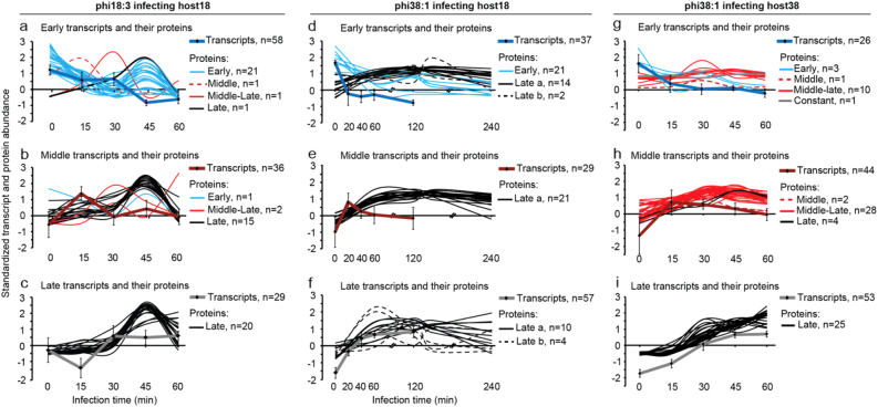
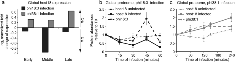
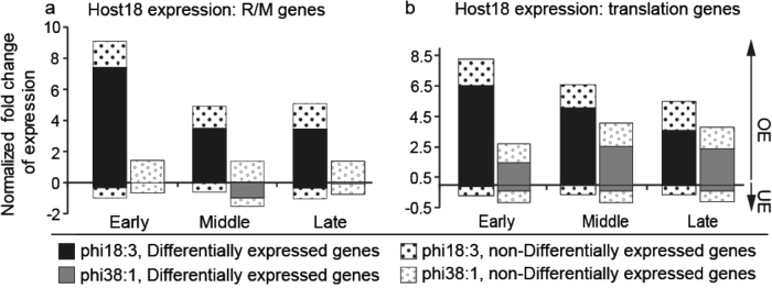
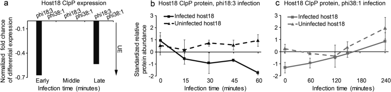
Phage-host interactions are critical to ecology, evolution, and biotechnology. Central to those is infection efficiency, which remains poorly understood, particularly in nature. Here we apply genome-wide transcriptomics and proteomics to investigate infection efficiency in nature's own experiment: two nearly identical (genetically and physiologically) Bacteroidetes bacterial strains (host18 and host38) that are genetically intractable, but environmentally important, where phage infection efficiency varies. On host18, specialist phage phi18:3 infects efficiently, whereas generalist phi38:1 infects inefficiently. On host38, only phi38:1 infects, and efficiently. Overall, phi18:3 globally repressed host18's transcriptome and proteome, expressed genes that likely evaded host restriction/modification (R/M) defenses and controlled its metabolism, and synchronized phage transcription with translation. In contrast, phi38:1 failed to repress host18's transcriptome and proteome, did not evade host R/M defenses or express genes for metabolism control, did not synchronize transcripts with proteins and its protein abundances were likely targeted by host proteases. However, on host38, phi38:1 globally repressed host transcriptome and proteome, synchronized phage transcription with translation, and infected host38 efficiently. Together these findings reveal multiple infection inefficiencies. While this contrasts the single mechanisms often revealed in laboratory mutant studies, it likely better reflects the phage-host interaction dynamics that occur in nature.
Conflict of interest statement
The authors declare that they have no conflict of interest.
Figures


Similar articles
-
Regulation of infection efficiency in a globally abundant marine Bacteriodetes virus.ISME J. 2017 Jan;11(1):284-295. doi: 10.1038/ismej.2016.81. Epub 2016 May 17. ISME J. 2017. PMID: 27187794 Free PMC article.
-
Contrasting genomic patterns and infection strategies of two co-existing Bacteroidetes podovirus genera.Environ Microbiol. 2014 Aug;16(8):2501-13. doi: 10.1111/1462-2920.12391. Epub 2014 Feb 18. Environ Microbiol. 2014. PMID: 24428166
-
Large-scale maps of variable infection efficiencies in aquatic Bacteroidetes phage-host model systems.Environ Microbiol. 2016 Nov;18(11):3949-3961. doi: 10.1111/1462-2920.13392. Epub 2016 Jun 27. Environ Microbiol. 2016. PMID: 27235779
-
Bacteriophage host range and bacterial resistance.Adv Appl Microbiol. 2010;70:217-48. doi: 10.1016/S0065-2164(10)70007-1. Epub 2010 Mar 6. Adv Appl Microbiol. 2010. PMID: 20359459 Review.
-
Molecular Basis of Bacterial Host Interactions by Gram-Positive Targeting Bacteriophages.Viruses. 2018 Jul 28;10(8):397. doi: 10.3390/v10080397. Viruses. 2018. PMID: 30060549 Free PMC article. Review.
Cited by
-
Genomic characterization of Pseudomonas syringae pv. syringae from Callery pear and the efficiency of associated phages in disease protection.Microbiol Spectr. 2024 Mar 5;12(3):e0283323. doi: 10.1128/spectrum.02833-23. Epub 2024 Feb 7. Microbiol Spectr. 2024. PMID: 38323825 Free PMC article.
-
Diversity and Host Interactions Among Virulent and Temperate Baltic Sea Flavobacterium Phages.Viruses. 2020 Jan 30;12(2):158. doi: 10.3390/v12020158. Viruses. 2020. PMID: 32019073 Free PMC article.
-
Phylogenomics and genetic analysis of solvent-producing Clostridium species.Sci Data. 2024 May 1;11(1):432. doi: 10.1038/s41597-024-03210-6. Sci Data. 2024. PMID: 38693191 Free PMC article.
-
Life and death in the soil microbiome: how ecological processes influence biogeochemistry.Nat Rev Microbiol. 2022 Jul;20(7):415-430. doi: 10.1038/s41579-022-00695-z. Epub 2022 Feb 28. Nat Rev Microbiol. 2022. PMID: 35228712 Review.
-
Ribosome profiling reveals downregulation of UMP biosynthesis as the major early response to phage infection.Microbiol Spectr. 2024 Apr 2;12(4):e0398923. doi: 10.1128/spectrum.03989-23. Epub 2024 Mar 7. Microbiol Spectr. 2024. PMID: 38451091 Free PMC article.
References
-
- Salmond GP, Fineran PC. A century of the phage: past, present and future. Nat Rev Microbiol. 2015;13:777–86. - PubMed
-
- Diaz-Munoz SL, Koskella B. Bacteria-phage interactions in natural environments. Adv Appl Microbiol. 2014;89:135–83. - PubMed
-
- Paez-Espino D, Eloe-Fadrosh EA, Pavlopoulos GA, Thomas AD, Huntemann M, Mikhailova N, et al. Uncovering Earth’s virome. Nature. 2016;536:425–30. - PubMed
Publication types
MeSH terms
Substances
Grants and funding
LinkOut - more resources
Full Text Sources
Other Literature Sources
Molecular Biology Databases
