

Serum autotaxin levels are correlated with hepatic fibrosis and ballooning in patients with non-alcoholic fatty liver disease
- PMID: 29568204
- PMCID: PMC5859226
- DOI: 10.3748/wjg.v24.i11.1239
Serum autotaxin levels are correlated with hepatic fibrosis and ballooning in patients with non-alcoholic fatty liver disease
Abstract
Aim: To examine the relationship between serum autotaxin (ATX) concentrations and clinicopathological findings in non-alcoholic fatty liver disease (NAFLD) patients.
Methods: One hundred eighty-six NAFLD patients who had undergone liver biopsy between 2008 and 2017 were retrospectively enrolled. Serum samples were collected at the time of biopsy and ATX was measured by enzyme immunoassays. Sera obtained from 160 healthy, non-obese individuals were used as controls. Histological findings were graded according to an NAFLD scoring system and correlations with serum ATX were calculated by Spearman's test. Diagnostic accuracy was evaluated using the area under the receiver operating characteristic curve (AUC). Cut-off values were identified by the Youden index, and the nearest clinically applicable value to the cutoff was considered the optimal threshold for clinical convenience.
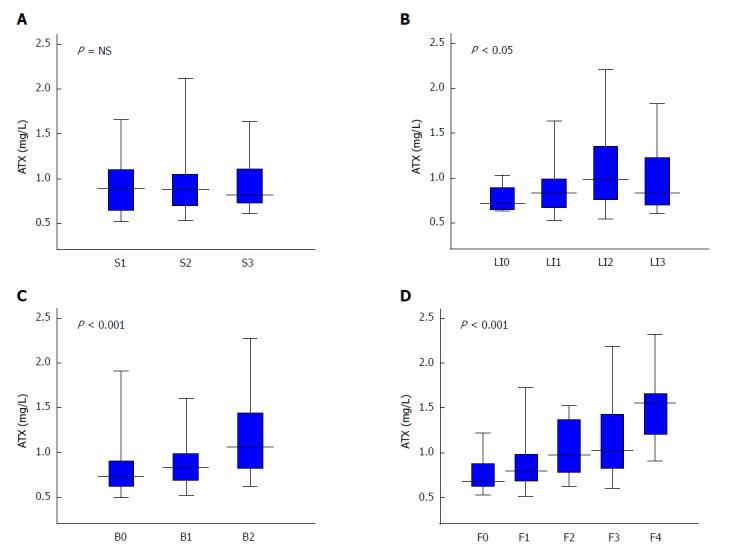
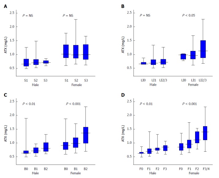
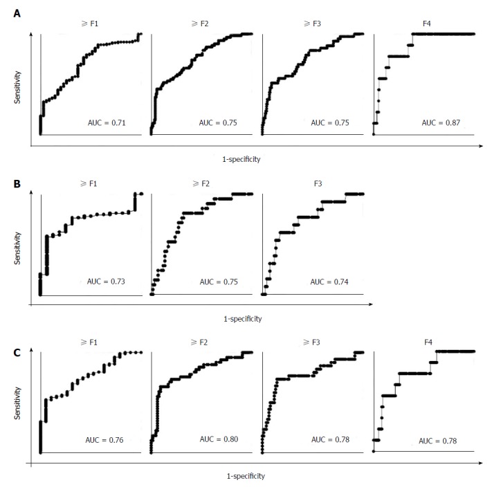
Results: Serum ATX levels were significantly higher in NAFLD patients than in controls (0.86 mg/L vs 0.76 mg/L, P < 0.001) and correlated significantly with ballooning score and fibrosis stage (r = 0.36, P < 0.001 and r = 0.45, P < 0.001, respectively). Such tendencies were stronger in female patients. There were no remarkable relationships between ATX and serum alanine aminotransferase, lipid profiles, or steatosis scores. The AUC values of ATX for predicting the presence of fibrosis (≥ F1), significant fibrosis (≥ F2), severe fibrosis (≥ F3), and cirrhosis (F4), were all more than 0.70 in respective analyses.
Conclusion: Serum ATX levels may at least partially reflect histological severity in NAFLD.
Keywords: Autotaxin; Ballooning; Fibrosis; Non-alcoholic fatty liver disease.
Conflict of interest statement
Conflict-of-interest statement: The authors declare that no conflict of interest exists.
Figures

Similar articles
-
Association of Serum Autotaxin Levels with Liver Fibrosis in Patients with Chronic Hepatitis C.Sci Rep. 2017 Apr 20;7:46705. doi: 10.1038/srep46705. Sci Rep. 2017. PMID: 28425454 Free PMC article.
-
Serum adipokines might predict liver histology findings in non-alcoholic fatty liver disease.World J Gastroenterol. 2016 Jun 7;22(21):5096-103. doi: 10.3748/wjg.v22.i21.5096. World J Gastroenterol. 2016. PMID: 27275102 Free PMC article.
-
Comparative diagnostic accuracy of red cell distribution width-to-platelet ratio versus noninvasive fibrosis scores for the diagnosis of liver fibrosis in biopsy-proven nonalcoholic fatty liver disease.Eur J Gastroenterol Hepatol. 2015 Nov;27(11):1293-9. doi: 10.1097/MEG.0000000000000445. Eur J Gastroenterol Hepatol. 2015. PMID: 26302023
-
Liver fibrosis in non-alcoholic fatty liver disease - diagnostic challenge with prognostic significance.World J Gastroenterol. 2015 Oct 21;21(39):11077-87. doi: 10.3748/wjg.v21.i39.11077. World J Gastroenterol. 2015. PMID: 26494963 Free PMC article. Review.
-
The independent effect of exercise on biopsy-proven non-alcoholic fatty liver disease: A systematic review.Clin Mol Hepatol. 2023 Feb;29(Suppl):S319-S332. doi: 10.3350/cmh.2022.0366. Epub 2022 Dec 14. Clin Mol Hepatol. 2023. PMID: 36517000 Free PMC article.
Cited by
-
Inhibition of autotaxin alleviates pathological features of hepatic encephalopathy at the level of gut-liver-brain axis: an experimental and bioinformatic study.Cell Death Dis. 2023 Aug 1;14(8):490. doi: 10.1038/s41419-023-06022-5. Cell Death Dis. 2023. PMID: 37528089 Free PMC article.
-
Role of autotaxin in systemic lupus erythematosus.Front Med (Lausanne). 2023 Apr 4;10:1166343. doi: 10.3389/fmed.2023.1166343. eCollection 2023. Front Med (Lausanne). 2023. PMID: 37122329 Free PMC article. Review.
-
Serum Autotaxin Levels Predict Liver-Related Events in Patients With Primary Biliary Cholangitis: A Long-Term Multicenter Observational Study.Clin Transl Gastroenterol. 2024 Dec 1;15(12):e00779. doi: 10.14309/ctg.0000000000000779. Clin Transl Gastroenterol. 2024. PMID: 39466702 Free PMC article.
-
Elevated Autotaxin and LPA Levels During Chronic Viral Hepatitis and Hepatocellular Carcinoma Associate with Systemic Immune Activation.Cancers (Basel). 2019 Nov 25;11(12):1867. doi: 10.3390/cancers11121867. Cancers (Basel). 2019. PMID: 31769428 Free PMC article. Review.
-
Effects of a 12-week whole-grain or refined wheat intervention on plasma acylcarnitines, bile acids and signaling lipids, and association with liver fat: A post-hoc metabolomics study of a randomized controlled trial.Front Nutr. 2022 Oct 13;9:1026213. doi: 10.3389/fnut.2022.1026213. eCollection 2022. Front Nutr. 2022. PMID: 36330140 Free PMC article.
References
MeSH terms
Substances
LinkOut - more resources
Full Text Sources
Other Literature Sources
Medical
Miscellaneous
