. 2018 Feb 3;9(17):13390-13406.
doi: 10.18632/oncotarget.24385. eCollection 2018 Mar 2.

Amiodarone promotes cancer cell death through elevated truncated SRSF3 and downregulation of miR-224

Affiliations

Amiodarone promotes cancer cell death through elevated truncated SRSF3 and downregulation of miR-224

Yung-Lung Chang et al. Oncotarget. .

Abstract

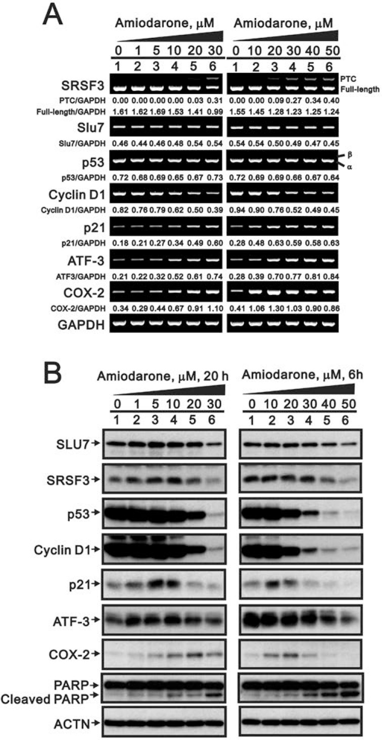
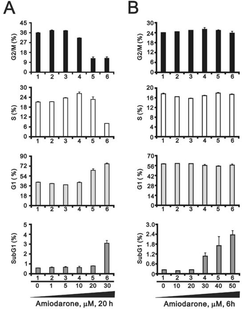
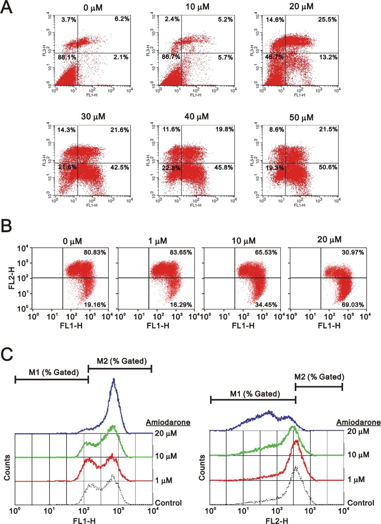
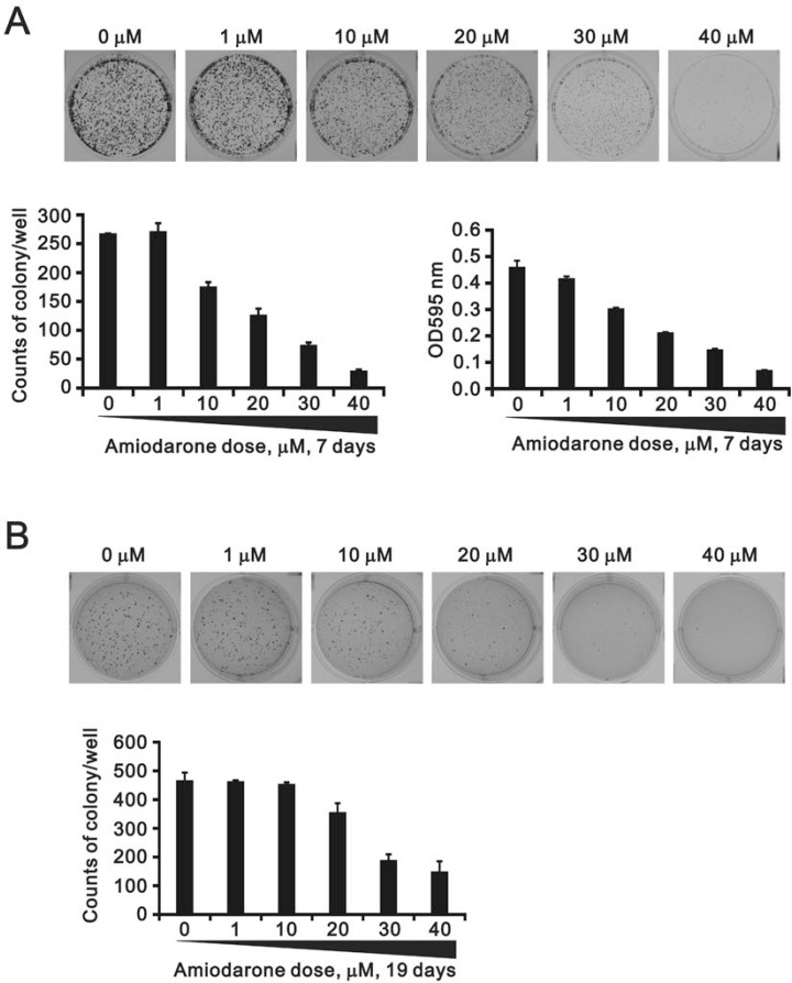
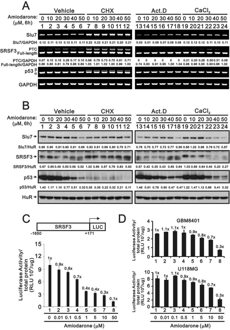
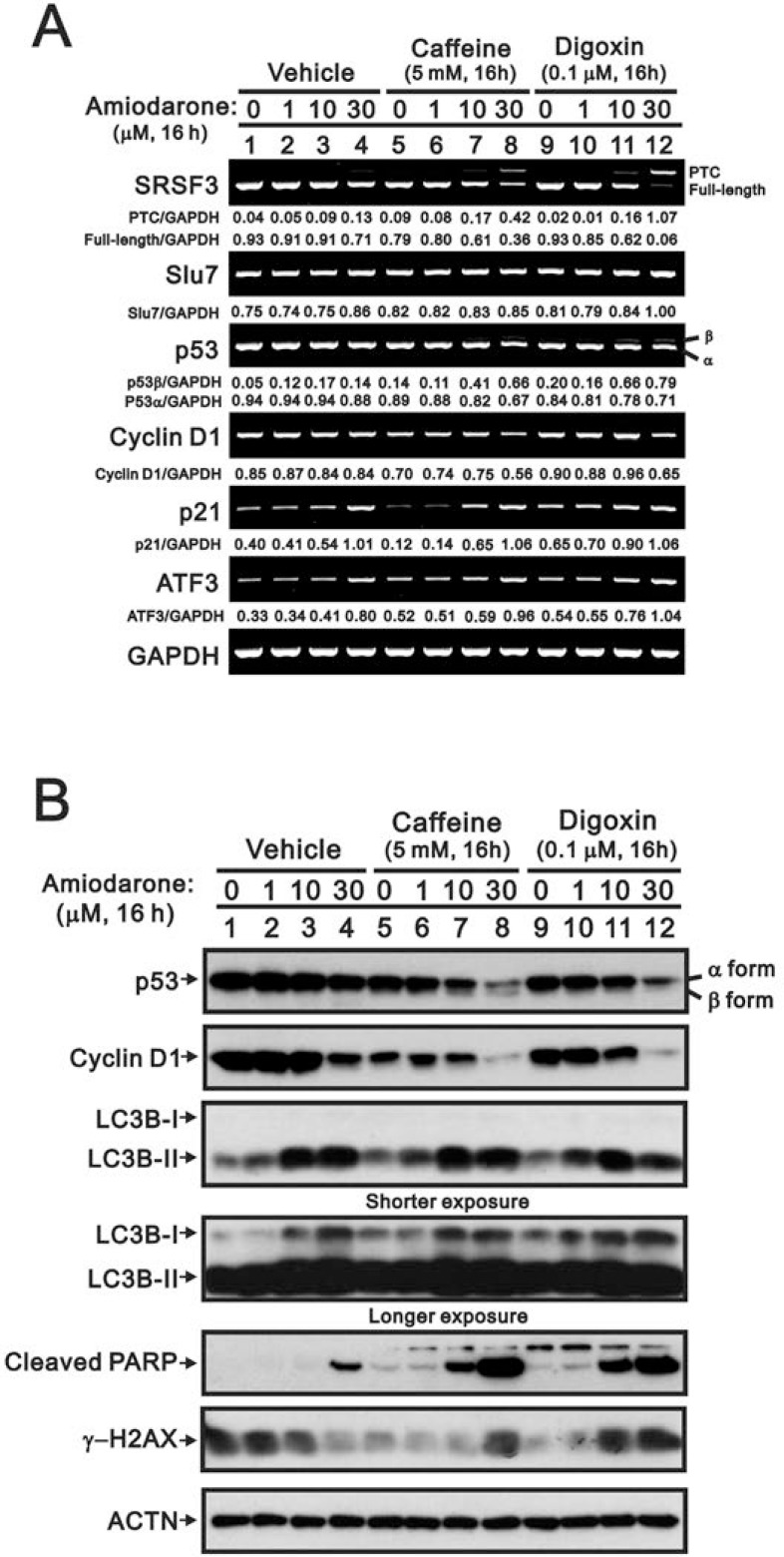
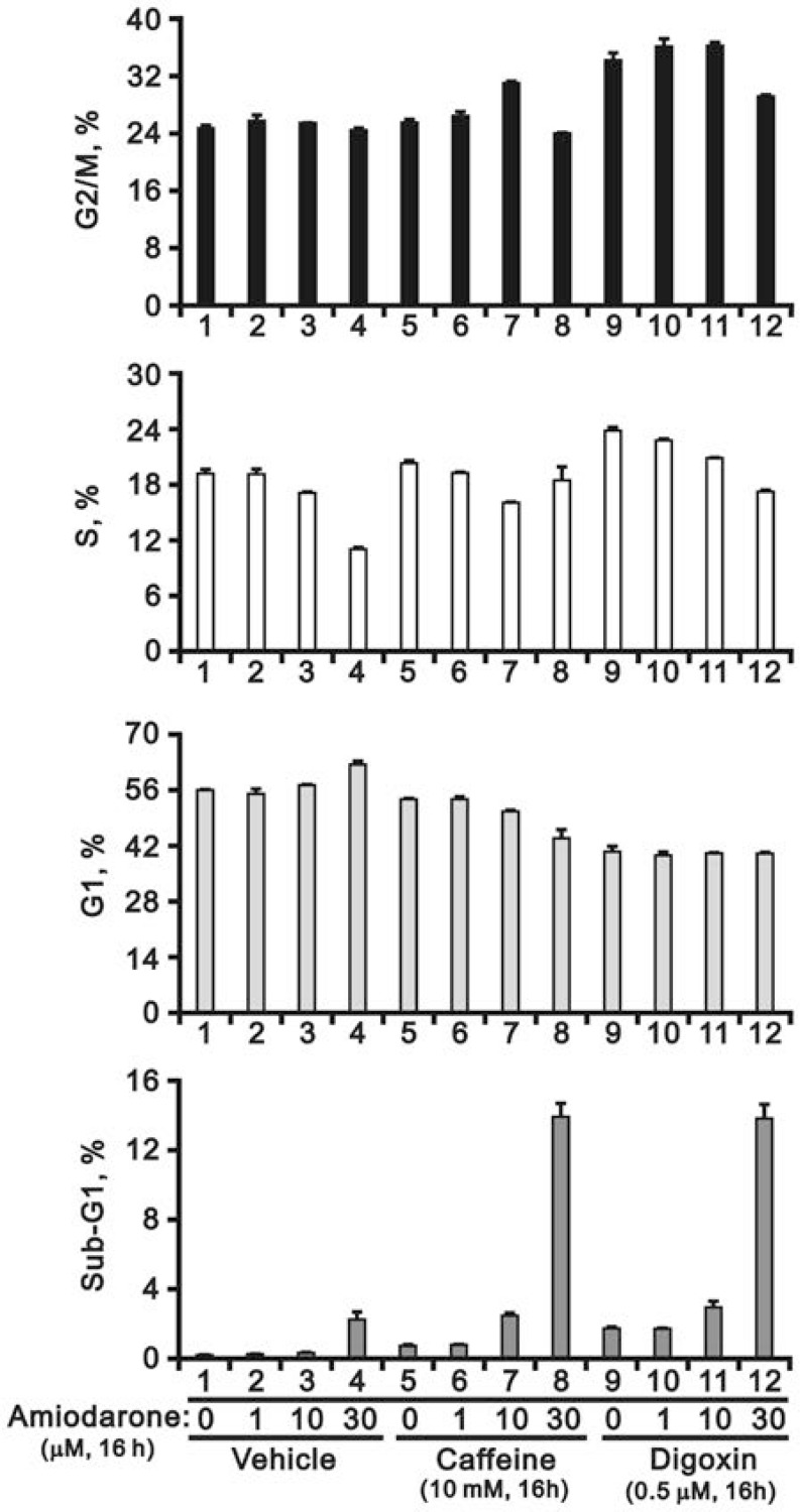
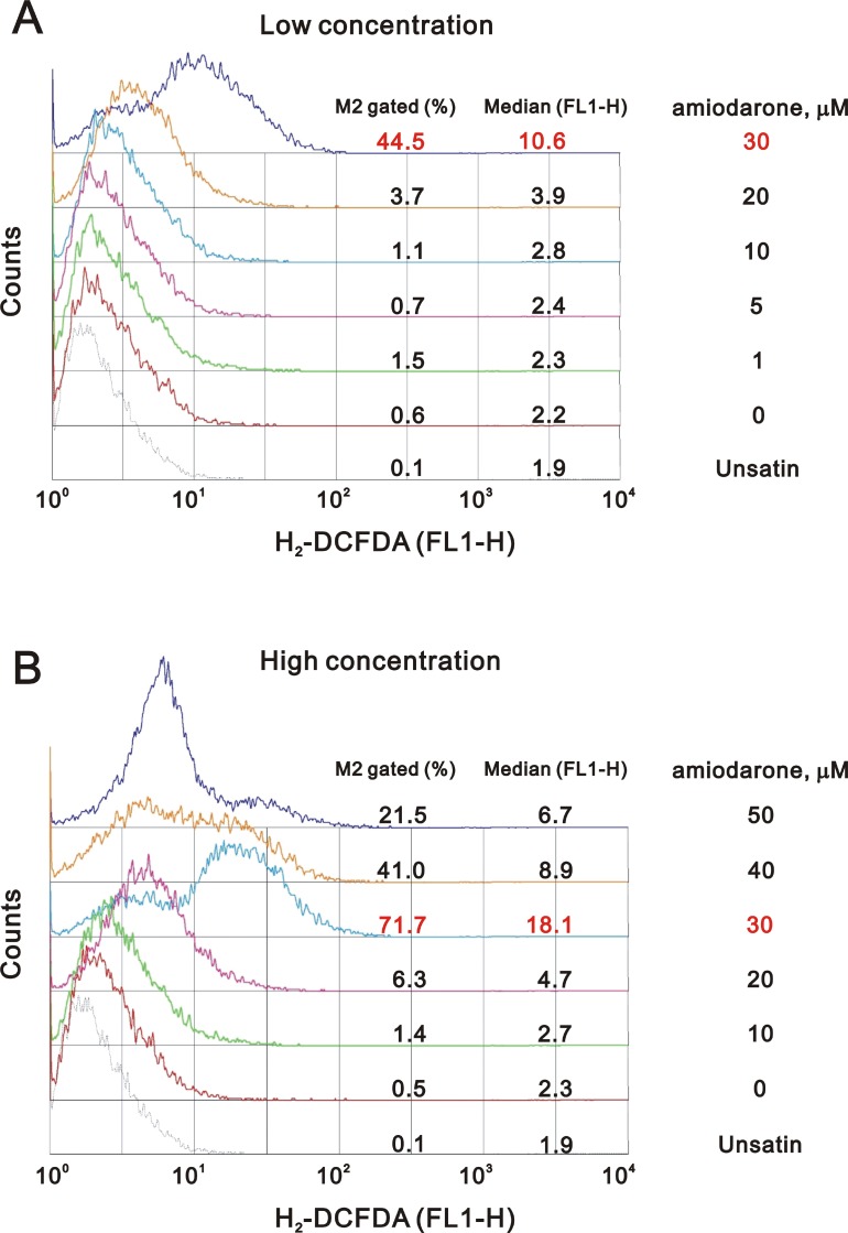
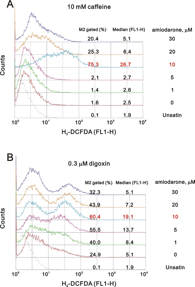
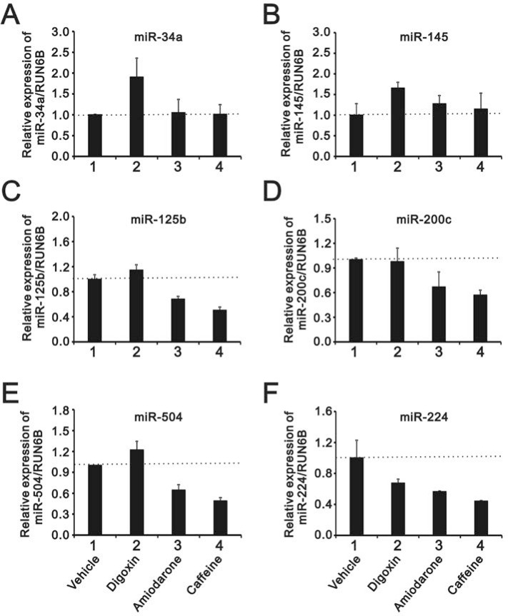
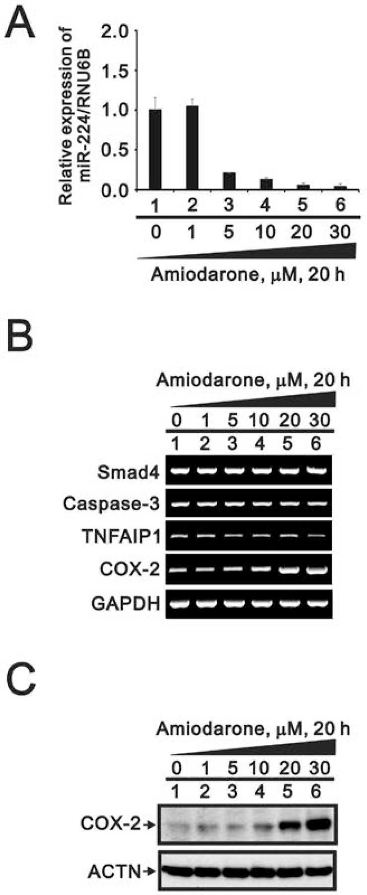
Amiodarone is a widely used class III antiarrhythmic agent which prolongs the action potential and refractory period by blockage of several types of myocardial potassium channels. Emerging evidence suggests that amiodarone sensitize tumor cells in response to chemotherapy. Nevertheless, little is known about the underlying molecular mechanism. To gain further insight, we demonstrated that amiodarone accumulated the population of a premature termination codon-containing isoform of serine and arginine rich splicing factor 3 (SRSF3-PTC) without increasing alternative spliced p53 beta isoform. Amiodarone enhanced reactive oxygen species production and increased cell apoptosis, whereas reduced DNA damage. Moreover, amiodarone suppressed miR-224 and increased its target COX-2 expression. Taken together, our results suggested amiodarone caused cancer cell death might be through increased SRSF3-PTC and miR-224 reduction in a p53-independent manner.

Keywords: amiodarone; autophagy; caffeine; digoxin; miR-224.


Conflict of interest statement

CONFLICTS OF INTEREST The authors declare no conflicts of interests related to this study.

Figures

Figure 1
Figure 1. The effects of amiodarone on target gene and protein expression in HeLa cells
HeLa cells were treated with indicated amount of amiodarone for indicated time. The cells were collected and subjected to (A) RT-PCR analysis of SRSF3, Slu7, p53, cyclin D1, p21, ATF3, COX-2, and GAPDH (loading control) mRNA expression; (B) immunoblot analysis for the detection of SLU7, SRSF3, p53, cyclin D1, p21, ATF3, COX-2, PARP, and ACTN (loading control) protein expression. PCR bands (A) were quantified through pixel density scanning and evaluated by ImageJ software, version 1.44a (http://imagej.nih.gov/ij/). The results are representative of two independent experiments.
Figure 2
Figure 2. Amiodarone caused cellular apoptosis
HeLa cells were treated with (A) lower concentration of amiodarone for 20 h and (B) higher concentration of amiodarone for 6 h. The cells were stained with PI, and the DNA content was determined by FACS analysis. The results are representative of three independent experiments.
Figure 3
Figure 3. Amiodarone triggered intrinsic apoptosis
HeLa cells were treated indicated amount amiodarone for 20 h. Cells were subject to the flow cytometry analysis using (A) the PE Annexin V Apoptosis Detection Kit (BD Biosciences) (B) the JC-1 staining kit. The results (A and B) are representative of three independent experiments. (C) The data from (B) were further measured the values of FL1-H and FL2-H.
Figure 4
Figure 4. The effects of amiodarone on the cell colony and anchorage-independent growth
(A) HeLa cells were treated indicated amount amiodarone for 7 days and cells were subject to the colony formation. After 7 days incubation, colonies were fixed with methanol and stained with 0.005% crystal violet solution for 1 h and photographed. Colonies were counted and quantified using ImageJ software. (B) HeLa cells were treated indicated amount amiodarone for 19 days and cells were subject to the colony formation in soft agar. After 19 days incubation, colonies were stained with 0.005% Crystal Violet solution for 30∼60 min and photographed. Colonies were counted and quantified using ImageJ software. All assays were done in triplicate.
Figure 5
Figure 5. The regulatory mechanism of amiodarone on the SRSF3 expression
HeLa cells were treated indicated amount amiodarone with pre-treated 2 h of vehicle, CHX (50 mg/ml), Act D (1 µM), and calcium chloride (1 mM) for 6 h. Cells were subject to the (A) Western blotting analysis and (B) RT-PCR analysis. The numerical data below each band is to indicate the ratio of specific mRNA (A) or protein (B) with control mRNA (GAPDH) or control protein (HuR) using the ImageJ software. The results (A and B) are representative of three independent experiments. (C and D) HeLa, GBM8401, U118MG cells were transiently transfected with 0.5 mg pGL3.SRSF3 (–1650/+171)-LUC and cells were treated with indicated amount of amiodarone for 20 h. Cells were harvested for luciferase reporter assay with the Promega Luciferase Assay Kit. The numbers (C and D) above the columns indicate the luciferase activity relative to an index of 1 for the reporter alone with vehicle.
Figure 6
Figure 6. Amiodarone synergistically enhanced caffeine- or digoxin-induced SRSF3 alternative splicing and p53-independent apoptosis in HeLa cells
HeLa cells were treated with 5 mM caffeine or 0.1 µM digoxin and along with indicated amount of amiodarone for 16 h. The cells were collected and subjected to (A) RT-PCR analysis of SRSF3, Slu7, p53, cyclin D1, p21, ATF3, COX-2, and GAPDH (loading control) mRNA expression; (B) immunoblot analysis for the detection of p53, cyclin D1, LC3B, PARP, γ−H2AX, and ACTN (loading control) protein expression. PCR bands (A) were quantified through pixel density scanning and evaluated by ImageJ software, version 1.44a (http://imagej.nih.gov/ij/). The results are representative of two independent experiments.
Figure 7
Figure 7. Amiodarone augmented the effects on the increase of sub-G1 population in HeLa cells by caffeine or digoxin
HeLa cells were treated with 5 mM caffeine or 0.1 µM digoxin and along with indicated amount of amiodarone for 16 h. The cells were stained with PI, and the DNA content was determined by FACS analysis. The results are representative of three independent experiments.
Figure 8
Figure 8. Amiodarone induced the ROS generation in HeLa cell
HeLa cells were treated with (A) lower concentration of amiodarone for 20 h and (B) higher concentration of amiodarone for 16 h. After incubated, we stained live cells with 10 µM DCFH-DA for 30∼60 min at 37° C, harvested cells were then subjected to FACS analysis. The results are representative of three independent experiments.
Figure 9
Figure 9. Caffeine or digoxin increased the effects of amiodarone for the elevation of ROS generation in HeLa cell
HeLa cells were treated with (A) 10 mM caffeine or (B) 0.3 µM digoxin and lower concentration of amiodarone for 16 h. After incubation, we stained live cells with 10 µM DCFH-DA for 30∼60 min at 37° C, harvested cells were then subjected to FACS analysis. The results are representative of three independent experiments.
Figure 10
Figure 10. The comparison of the effects of caffeine, amiodarone, and digoxin on the miRs regulation in HeLa cell
HeLa cells were treated with 10 mM caffeine, 0.3 µM digoxin, and 30 mM amiodarone for 16 h. The cells were collected and subjected to the miR analysis for (A) miR-34a; (B) miR-145; (C) miR-125b; (D) miR-200c; (E) miR-504; and (F) miR-224 expression. The results are representative of three independent experiments.
Figure 11
Figure 11. The effects of amiodarone on miR-224 and its target genes in HeLa cell
HeLa cells were treated with lower concentration of amiodarone for 20 h. The cells were collected and subjected to (A) the miR analysis for miR-224 expression; (B) the RT-PCR analysis for caspase3, TNFAIP1, COX-2, and GAPDH (loading control); and (C) the Western blotting analysis using antibodies against COX-2 and ACTN (loading control). The results are representative of three independent experiments.

References

    1. Ellison KE, Stevenson WG, Sweeney MO, Epstein LM, Maisel WH. Management of arrhythmias in heart failure. Congest Heart Fail. 2003;9:91–99. - PubMed
    1. Singh BN. Current antiarrhythmic drugs: an overview of mechanisms of action and potential clinical utility. J Cardiovasc Electrophysiol. 1999;10:283–301. - PubMed
    1. Singh BN. Amiodarone as paradigm for developing new drugs for atrial fibrillation. J Cardiovasc Pharmacol. 2008;52:300–305. - PubMed
    1. Kathofer S, Thomas D, Karle CA. The novel antiarrhythmic drug dronedarone: comparison with amiodarone. Cardiovasc Drug Rev. 2005;23:217–230. - PubMed
    1. Karavelioglu Y, Karapinar H, Yuksel M, Memic K, Sarak T, Kurt R, Yilmaz A. Neutrophil to lymphocyte ratio is predictor of atrial fibrillation recurrence after cardioversion with amiodarone. Clin Appl Thromb Hemost. 2015;21:5–9. - PubMed