

Effects of Cetuximab and Erlotinib on the behaviour of cancer stem cells in head and neck squamous cell carcinoma
- PMID: 29568372
- PMCID: PMC5862593
- DOI: 10.18632/oncotarget.24416
Effects of Cetuximab and Erlotinib on the behaviour of cancer stem cells in head and neck squamous cell carcinoma
Abstract
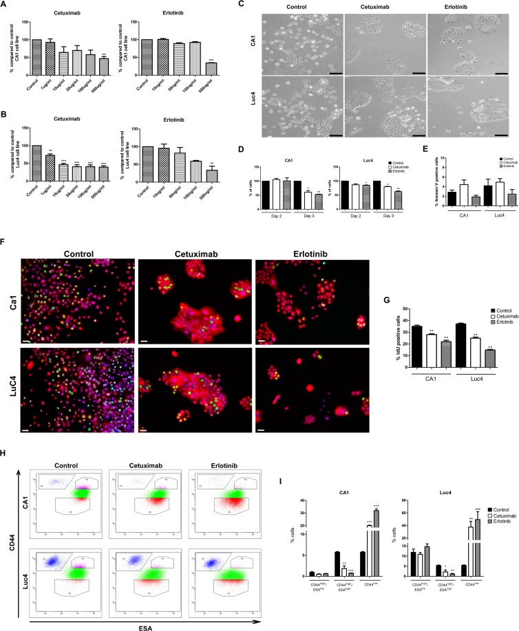
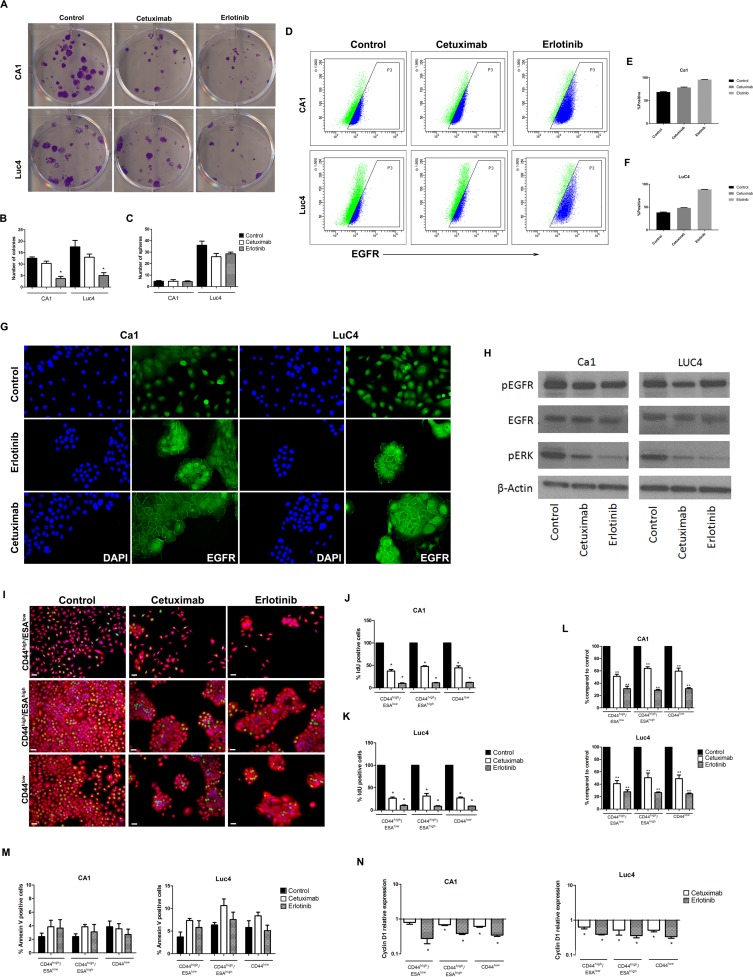
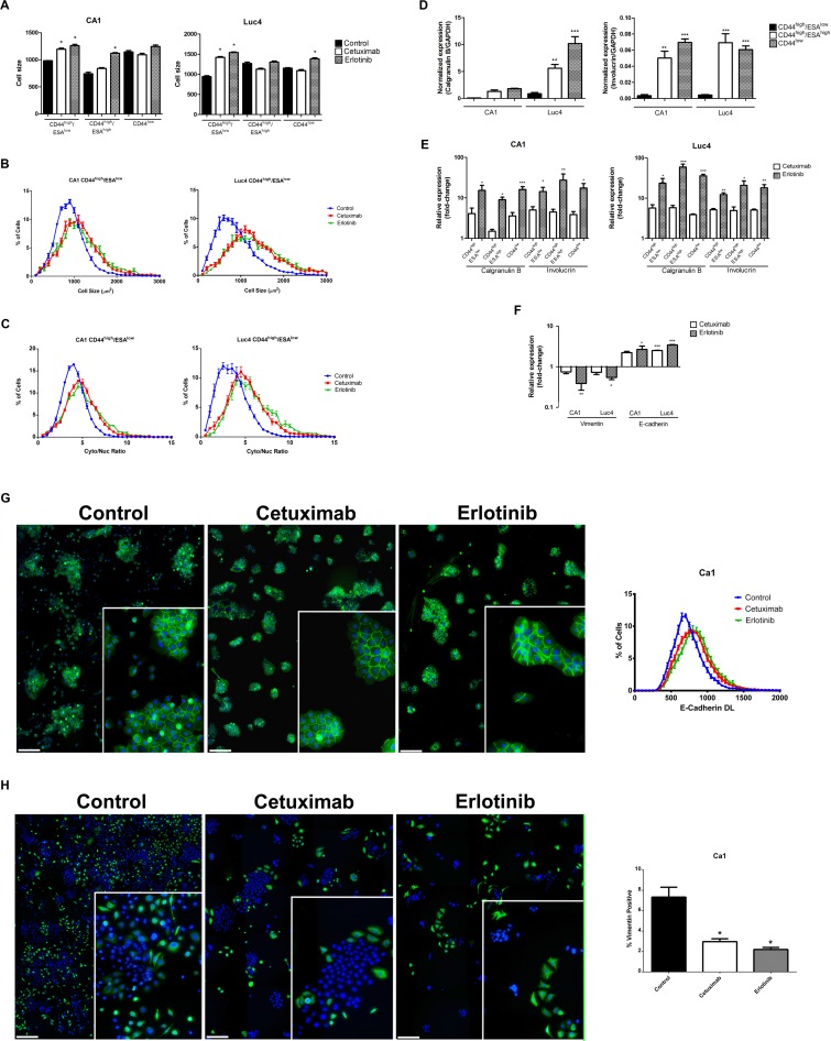
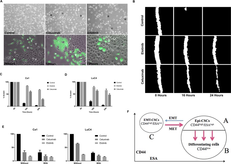
The therapeutic responses of many solid tumours to chemo- and radio-therapies are far from fully effective but therapies targeting malignancy-related cellular changes show promise for further control. In head and neck squamous cell carcinoma, the epidermal growth factor receptor (EGFR) is commonly overexpressed and investigation of agents that block this receptor indicate a limited response when used alone but an ability to enhance the actions of other drugs. The hierarchical stem cell patterns present in tumours generate cellular heterogeneity and this is further complicated by cancer stem cells (CSC) shifting between epithelial (Epi-CSC) and mesenchymal (EMT-CSC) states. To clarify how such heterogeneity influences responses to EGFR blocking, we examined the effects of Cetuximab and Erlotinib on the cell sub-populations in HNSCC cell lines. These agents reduced cell proliferation for all subpopulations but induced little cell death. They did however induce large shifts of cells between the EMT-CSC, Epi-CSC and differentiating cell compartments. Loss of EMT-CSCs reduced cell motility and is expected to reduce invasion and metastasis. EGFR blocking also induced shifts of Epi-CSCs into the differentiating cell compartment which typically has greater sensitivity to chemo/radiation, an effect expected to enhance the overall response of tumour cell populations to adjunctive therapies.
Keywords: Cetuximab; EMT; HNSCC; cancer stem cells; differentiation.
Conflict of interest statement
CONFLICTS OF INTEREST The authors declare no competing financial interests.
Figures
Similar articles
-
Afatinib radiosensitizes head and neck squamous cell carcinoma cells by targeting cancer stem cells.Oncotarget. 2017 Mar 28;8(13):20961-20973. doi: 10.18632/oncotarget.15468. Oncotarget. 2017. PMID: 28423495 Free PMC article.
-
Epithelial-to-mesenchymal transition and cancer stem(-like) cells in head and neck squamous cell carcinoma.Cancer Lett. 2013 Sep 10;338(1):47-56. doi: 10.1016/j.canlet.2012.06.013. Epub 2012 Jul 4. Cancer Lett. 2013. PMID: 22771535 Review.
-
Dual-agent molecular targeting of the epidermal growth factor receptor (EGFR): combining anti-EGFR antibody with tyrosine kinase inhibitor.Cancer Res. 2004 Aug 1;64(15):5355-62. doi: 10.1158/0008-5472.CAN-04-0562. Cancer Res. 2004. PMID: 15289342
-
Snail-induced epithelial-mesenchymal transition promotes cancer stem cell-like phenotype in head and neck cancer cells.Int J Oncol. 2014 Mar;44(3):693-9. doi: 10.3892/ijo.2013.2225. Epub 2013 Dec 23. Int J Oncol. 2014. PMID: 24365974
-
Induction of metastasis, cancer stem cell phenotype, and oncogenic metabolism in cancer cells by ionizing radiation.Mol Cancer. 2017 Jan 30;16(1):10. doi: 10.1186/s12943-016-0577-4. Mol Cancer. 2017. PMID: 28137309 Free PMC article. Review.
Cited by
-
Therapeutic Targeting of Cancer Stem Cells in Lung, Head and Neck, and Bladder Cancers.Cancers (Basel). 2021 Oct 12;13(20):5098. doi: 10.3390/cancers13205098. Cancers (Basel). 2021. PMID: 34680249 Free PMC article. Review.
-
Tumor microenvironment enriches the stemness features: the architectural event of therapy resistance and metastasis.Mol Cancer. 2022 Dec 22;21(1):225. doi: 10.1186/s12943-022-01682-x. Mol Cancer. 2022. PMID: 36550571 Free PMC article. Review.
-
Oral Cancer Stem Cells: Therapeutic Implications and Challenges.Front Oral Health. 2021 Jul 21;2:685236. doi: 10.3389/froh.2021.685236. eCollection 2021. Front Oral Health. 2021. PMID: 35048028 Free PMC article. Review.
-
Molecular Markers of Anticancer Drug Resistance in Head and Neck Squamous Cell Carcinoma: A Literature Review.Cancers (Basel). 2018 Oct 10;10(10):376. doi: 10.3390/cancers10100376. Cancers (Basel). 2018. PMID: 30308958 Free PMC article. Review.
-
Dual EGFR blockade with cetuximab and erlotinib combined with anti-VEGF antibody bevacizumab in advanced solid tumors: a phase 1 dose escalation triplet combination trial.Exp Hematol Oncol. 2020 Apr 20;9:7. doi: 10.1186/s40164-020-00159-1. eCollection 2020. Exp Hematol Oncol. 2020. PMID: 32337094 Free PMC article.
References
-
- Jemal A, Bray F, Center MM, Ferlay J, Ward E, Forman D. Global cancer statistics. CA Cancer J Clin. 2011;61:69–90. - PubMed
-
- Haddad RI, Shin DM. Recent advances in head and neck cancer. N Engl J Med. 2008;359:1143–1154. - PubMed
-
- Siegel RL, Miller KD, Jemal A. Cancer Statistics, 2017. CA Cancer J Clin. 2017;67:7–30. - PubMed
-
- Vermorken JB, Specenier P. Optimal treatment for recurrent/metastatic head and neck cancer. Ann Oncol. 2010;21:vii252–261. - PubMed
LinkOut - more resources
Full Text Sources
Other Literature Sources
Research Materials
Miscellaneous
