

Toward a patient-specific tissue engineered vascular graft
- PMID: 29568478
- PMCID: PMC5858675
- DOI: 10.1177/2041731418764709
Toward a patient-specific tissue engineered vascular graft
Abstract
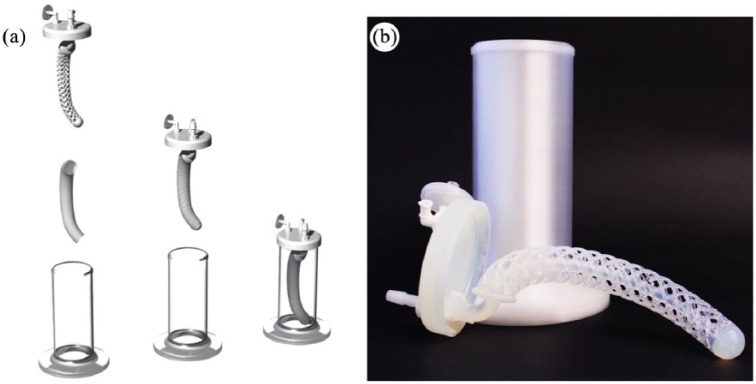
Integrating three-dimensional printing with the creation of tissue-engineered vascular grafts could provide a readily available, patient-specific, autologous tissue source that could significantly improve outcomes in newborns with congenital heart disease. Here, we present the recent case of a candidate for our tissue-engineered vascular graft clinical trial deemed ineligible due to complex anatomical requirements and consider the application of three-dimensional printing technologies for a patient-specific graft. We 3D-printed a closed-disposable seeding device and validated that it performed equivalently to the traditional open seeding technique using ovine bone marrow-derived mononuclear cells. Next, our candidate's preoperative imaging was reviewed to propose a patient-specific graft. A seeding apparatus was then designed to accommodate the custom graft and 3D-printed on a commodity fused deposition modeler. This exploratory feasibility study represents an important proof of concept advancing progress toward a rationally designed patient-specific tissue-engineered vascular graft for clinical application.
Keywords: 3D-printing; Fontan operation; Tissue-engineered vascular graft; cell seeding; patient-specific modeling.
Conflict of interest statement
Declaration of conflicting interests: The author(s) declared the following potential conflicts of interest with respect to the research, authorship, and/or publication of this article: C.Breuer is on the Scientific Advisory board of Cook Medical (Bloomington, IN), and C.Breuer and T.S. received research support from Gunze, Ltd (Kyoto, Japan) and Cook Regentec (Indianapolis, IN). Gunze, Ltd. kindly provided the scaffolds used in this study. C. Breuer and C. Best are cofounders of LYST Therapeutics, LLC (Columbus, OH). The remaining authors have no conflicts of interest to disclose.
Figures


Similar articles
-
Preclinical study of patient-specific cell-free nanofiber tissue-engineered vascular grafts using 3-dimensional printing in a sheep model.J Thorac Cardiovasc Surg. 2017 Apr;153(4):924-932. doi: 10.1016/j.jtcvs.2016.10.066. Epub 2016 Nov 14. J Thorac Cardiovasc Surg. 2017. PMID: 27938900 Free PMC article.
-
Construction of an autologous tissue-engineered venous conduit from bone marrow-derived vascular cells: optimization of cell harvest and seeding techniques.J Pediatr Surg. 2007 Jan;42(1):198-202. doi: 10.1016/j.jpedsurg.2006.09.054. J Pediatr Surg. 2007. PMID: 17208565
-
Designing a tissue-engineered tracheal scaffold for preclinical evaluation.Int J Pediatr Otorhinolaryngol. 2018 Jan;104:155-160. doi: 10.1016/j.ijporl.2017.10.036. Epub 2017 Nov 22. Int J Pediatr Otorhinolaryngol. 2018. PMID: 29287858 Free PMC article.
-
Tissue engineered vascular grafts for pediatric cardiac surgery.Transl Pediatr. 2018 Apr;7(2):188-195. doi: 10.21037/tp.2018.02.01. Transl Pediatr. 2018. PMID: 29770300 Free PMC article. Review.
-
Three-dimensional (3D) printed scaffold and material selection for bone repair.Acta Biomater. 2019 Jan 15;84:16-33. doi: 10.1016/j.actbio.2018.11.039. Epub 2018 Nov 24. Acta Biomater. 2019. PMID: 30481607 Review.
Cited by
-
Small-Diameter Blood Vessel Substitutes: Biomimetic Approaches to Improve Patency.Biomimetics (Basel). 2024 Feb 7;9(2):97. doi: 10.3390/biomimetics9020097. Biomimetics (Basel). 2024. PMID: 38392143 Free PMC article. Review.
-
Biological Scaffolds for Congenital Heart Disease.Bioengineering (Basel). 2023 Jan 2;10(1):57. doi: 10.3390/bioengineering10010057. Bioengineering (Basel). 2023. PMID: 36671629 Free PMC article. Review.
-
Aortic "Disease-in-a-Dish": Mechanistic Insights and Drug Development Using iPSC-Based Disease Modeling.Front Cell Dev Biol. 2020 Oct 28;8:550504. doi: 10.3389/fcell.2020.550504. eCollection 2020. Front Cell Dev Biol. 2020. PMID: 33195187 Free PMC article. Review.
-
Graft rejection in paediatric congenital heart disease.Transl Pediatr. 2023 Aug 30;12(8):1572-1591. doi: 10.21037/tp-23-80. Epub 2023 Aug 21. Transl Pediatr. 2023. PMID: 37692547 Free PMC article. Review.
-
Implanted Tissue-Engineered Vascular Graft Cell Isolation with Single-Cell RNA Sequencing Analysis.Tissue Eng Part C Methods. 2023 Feb;29(2):72-84. doi: 10.1089/ten.TEC.2022.0189. Tissue Eng Part C Methods. 2023. PMID: 36719780 Free PMC article.
References
-
- Hoffman JIE, Kaplan S. The incidence of congenital heart disease. J Am Coll Cardiol 2002; 39: 1890–1900. - PubMed
-
- Rychik J, Goldberg D, Rand E, et al. End-organ consequences of the Fontan operation: liver fibrosis, protein-losing enteropathy and plastic bronchitis. Cardiol Young 2013; 23: 831–840. - PubMed
-
- Binns RL, Ku DN, Stewart MT, et al. Optimal graft diameter: effect of wall shear stress on vascular healing. J Vasc Surg 1989; 10: 326–337. - PubMed
Grants and funding
LinkOut - more resources
Full Text Sources
Other Literature Sources
