Review
. 2018 Mar 12:7:307.
doi: 10.12688/f1000research.13179.1. eCollection 2018.

Renal cell carcinoma: a review of biology and pathophysiology

Affiliations
Review

Renal cell carcinoma: a review of biology and pathophysiology

Shahzaib Nabi et al. F1000Res. .

Abstract

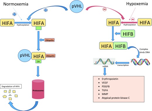
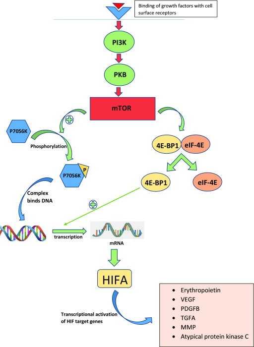
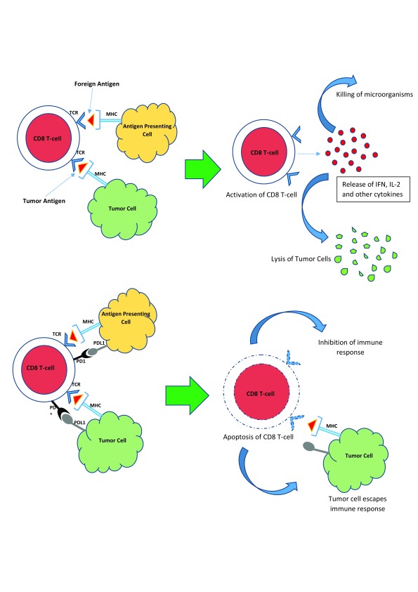
Over the past decade, our understanding of the biology and pathophysiology of renal cell carcinoma (RCC) has improved significantly. Insight into the disease process has helped us in developing newer therapeutic approaches toward RCC. In this article, we review the various genetic and immune-related mechanisms involved in the pathogenesis and development of this cancer and how that knowledge is being used to develop therapeutic targeted drugs for the treatment of RCC. The main emphasis of this review article is on the most common genetic alterations found in clear cell RCC and how various drugs are currently targeting such pathways. This article also looks at the role of the immune system in allowing the growth of RCC and how the immune system can be manipulated to reactivate cytotoxic immunity against RCC.

Keywords: BAP-1; Glutaminase; HIF; Immunotherapy; PBRM-1; VHL; biology; mTOR; renal cell carcinoma; von Hippel Lindau.


Conflict of interest statement

Competing interests: ETL receives kidney cancer clinical trial funding from Argos, Bristol-Myers Squibb, Calithera, Genentech, Merck, Peloton, Pfizer, and Roche; has served as a consultant for Bristol-Myers Squibb; and serves on the Independent Data Monitoring Board for Calithera. ERK receives kidney cancer clinical trial funding from Bristol-Myers Squibb. TWF receives clinical trial funding from Bristol-Myers Squibb, Novartis, Pfizer, Bavarian Nordic, Cougar Biotechnology, Dendreon, GTx, Janssen Oncology, Medivation, Sanofi, Genentech, Roche, Exelixis, Aragon Pharmaceuticals, Sotio, Tokai Pharmaceuticals, Astra-Zeneca/MedImmune, Lilly, Astellas, Agensys, and Seattle Genetics and has served as a consultant for GTx.No competing interests were disclosed.No competing interests were disclosed.

Figures

Figure 1.
Figure 1.. VHL/HIF axis.
Under normal oxygen tension, HIF1A and HIF2A are hydroxylated on proline residues and bind the pVHL, resulting in the polyubiquitination of HIFA, which targets it for proteasomal degradation. Under conditions of hypoxemia or in the absence of pVHL, hydroxylation of HIF1A and HIF2A does not occur and HIFA accumulates and dimerizes with HIFB and acts as a transcription factor, resulting in increased mRNA levels coding for VEGF, PDGFB, TGFA, erythropoietin, and extracellular matrix protein. HIFA, hypoxia-inducible factor alpha; HIFB, hypoxia-inducible factor beta; MMP, matrix metalloproteinase protein; mRNA, messenger RNA; PDGFB, platelet-derived growth factor beta; pVHL, protein of Von Hippel–Lindau gene; TGFA, transforming growth factor alpha; VEGF, vascular endothelial growth factor.
Figure 2.
Figure 2.. The mTOR–PI3K pathway.
Binding of cell surface growth factors activates phosphatidylinositol-3-kinase (PI3K) protein, which in turn activates mTOR, creating mTOR complexes 1 and 2 (mTORC1 and mTORC2), resulting in the phosphorylation of P70S6K and 4E-BP1/eIF4E and increased production of angiogenic proteins such as VEGF, PDGF, and TGFB, leading to cell growth and tumor progression. 4E-BP1, 4E-binding protein 1; eIF-4E, eukaryotic initiation factor-4E; HIF, hypoxia-inducible factor; HIFA, hypoxia-inducible factor alpha; MMP, matrix metalloproteinase; mRNA, messenger RNA; mTOR, mammalian target of rapamycin; PDGFB, platelet-derived growth factor beta; PI3K, phosphatidylinositol-3-kinase; PKB, protein kinase B; TGFA, transforming growth factor alpha; VEGF, vascular endothelial growth factor.
Figure 3.
Figure 3.. The programmed death-1 receptor pathway.
Recognition of foreign or tumor antigens normally results in the activation of CD8 T cells, leading to the killing of viruses or bacteria or the lysis of tumor cells. The programmed death-1 (PD-1) receptor is a cell membrane protein present on the surface of CD8 T cells, which, upon interacting with PD-L1 and PD-L2 on the surface of tumor cells, results in the suppression of the cytotoxic immune response, leading to tumor escape.IFN, interferon; IL-2, interleukin-2; MHC, major histocompatibility complex; PD-1, programmed death-1; PD-L1, programmed death ligand-1; TCR, T-cell receptor.

References

    1. Hsieh JJ, Purdue MP, Signoretti S, et al. : Renal cell carcinoma. Nat Rev Dis Primers. 2017;3:17009. 10.1038/nrdp.2017.9 - DOI - PMC - PubMed
    2. F1000 Recommendation

    1. Chow WH, Dong LM, Devesa SS: Epidemiology and risk factors for kidney cancer. Nat Rev Urol. 2010;7(5):245–57. 10.1038/nrurol.2010.46 - DOI - PMC - PubMed
    1. Siegel RL, Miller KD, Jemal A: Cancer Statistics, 2017. CA Cancer J Clin. 2017;67(1):7–30. 10.3322/caac.21387 - DOI - PubMed
    2. F1000 Recommendation

    1. Thompson RH, Ordonez MA, Iasonos A, et al. : Renal cell carcinoma in young and old patients--is there a difference? J Urol. 2008;180(4):1262–6; discussion 1266. 10.1016/j.juro.2008.06.037 - DOI - PMC - PubMed
    1. Siegel R, Ward E, Brawley O, et al. : Cancer statistics, 2011: the impact of eliminating socioeconomic and racial disparities on premature cancer deaths. CA Cancer J Clin. 2011;61(4):212–36. 10.3322/caac.20121 - DOI - PubMed