

Both activated and less-activated regions identified by functional MRI reconfigure to support task executions
- PMID: 29568689
- PMCID: PMC5853621
- DOI: 10.1002/brb3.893
Both activated and less-activated regions identified by functional MRI reconfigure to support task executions
Abstract
Introduction: Functional magnetic resonance imaging (fMRI) has become very important for noninvasively characterizing BOLD signal fluctuations, which reflect the changes in neuronal firings in the brain. Unlike the activation detection strategy utilized with fMRI, which only emphasizes the synchronicity between the functional nodes (activated regions) and the task design, brain connectivity and network theory are able to decipher the interactive structure across the entire brain. However, little is known about whether and how the activated/less-activated interactions are associated with the functional changes that occur when the brain changes from the resting state to a task state. What are the key networks that play important roles in the brain state changes?
Methods: We used the fMRI data from the Human Connectome Project S500 release to examine the changes of network efficiency, interaction strength, and fractional modularity contributions of both the local and global networks, when the subjects change from the resting state to seven different task states.
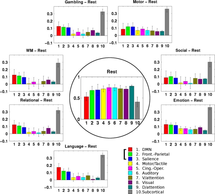
Results: We found that, from the resting state to each of seven task states, both the activated and less-activated regions had significantly changed to be in line with, and comparably contributed to, a global network reconfiguration. We also found that three networks, the default mode network, frontoparietal network, and salience network, dominated the flexible reconfiguration of the brain.
Conclusions: This study shows quantitatively that contributions from both activated and less-activated regions enable the global functional network to respond when the brain switches from the resting state to a task state and suggests the necessity of considering large-scale networks (rather than only activated regions) when investigating brain functions in imaging cognitive neuroscience.
Keywords: activation; brain network; functional connectivity; functional magnetic resonance imaging; network reconfiguration.
Figures


References
-
- Achard, S. , & Bullmore, E. (2007). Efficiency and cost of economical brain functional networks. PLoS computational biology, 3, e17 https://doi.org/10.1371/journal.pcbi.0030017 - DOI - PMC - PubMed
-
- Barch, D. M. , Burgess, G. C. , Harms, M. P. , Petersen, S. E. , Schlaggar, B. L. , Corbetta, M. , … Consortium, W. U.‐M. H. (2013). Function in the human connectome: Task‐fMRI and individual differences in behavior. NeuroImage, 80, 169–89. https://doi.org/10.1016/j.neuroimage.2013.05.033 - DOI - PMC - PubMed
-
- Bassett, D. S. , Porter, M. A. , Wymbs, N. F. , Grafton, S. T. , Carlson, J. M. , & Mucha, P. J. (2013). Robust detection of dynamic community structure in networks. Chaos, 23, 013142 https://doi.org/10.1063/1.4790830 - DOI - PMC - PubMed
-
- Bassett, D. S. , Yang, M. , Wymbs, N. F. , & Grafton, S. T. (2015). Learning‐induced autonomy of sensorimotor systems. Nature Neuroscience, 18, 744–751. https://doi.org/10.1038/nn.3993 - DOI - PMC - PubMed
-
- Beckmann, C. F. , Jenkinson, M. , & Smith, S. M. (2003). General multilevel linear modeling for group analysis in FMRI. NeuroImage, 20, 1052–1063. https://doi.org/10.1016/S1053-8119(03)00435-X - DOI - PubMed
Publication types
MeSH terms
Grants and funding
LinkOut - more resources
Full Text Sources
Other Literature Sources
Medical
