. 2018 Feb 7:2018:8497283.
doi: 10.1155/2018/8497283. eCollection 2018.

Experimental Nanopulse Ablation of Multiple Membrane Parasite on Ex Vivo Hydatid Cyst

Affiliations

Experimental Nanopulse Ablation of Multiple Membrane Parasite on Ex Vivo Hydatid Cyst

Xinhua Chen et al. Biomed Res Int. .

Abstract

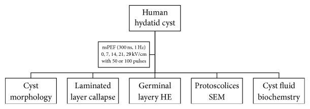
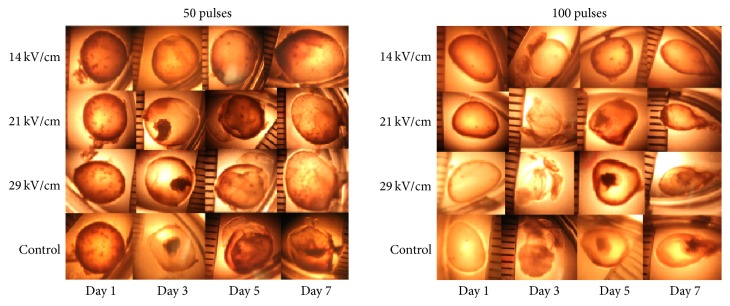
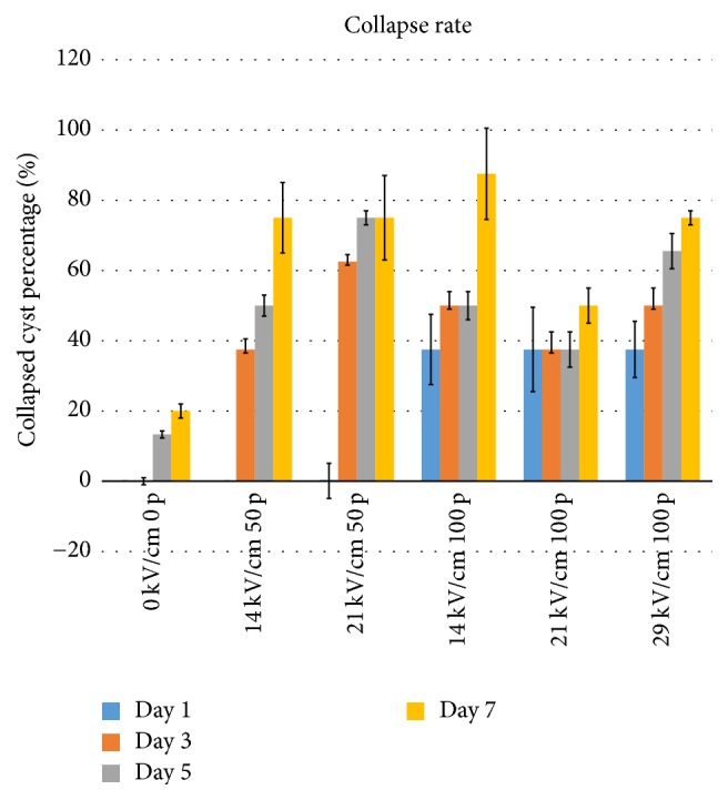
The impact of ultrashort nanopulse on cellular membrane is of biological significance and thus has been studied intensively. Different from cell study, this ex vivo study aims to investigate the biological effects of nanosecond pulsed electric field (nsPEF) on an independent multimembrane parasite, human hydatid cyst, to observe the unique influence of nanopulse on macromembrane structure, permeabilization, and biochemistry. The 300 ns nsPEF was delivered on an experimental model of single human hydatid cyst ex vivo with eight different parameters. Then pathological changes during 7 days of 48 parasite cysts were followed up after nsPEF. The laminated layer, the germinal layer, the protoscolex, and cyst fluid were evaluated by the morphological, pathological, and biochemical measurements. The parameter screening found that nsPEF can damage hydatid cyst effectively when the field strength is higher than 14 kV/cm. When nsPEF is higher than 29 kV/cm, nsPEF destroy hydatid cyst completely by collapsing the germinal layer, destructing protoscolices, and exhausting the nutrition.


Figures

Figure 1
Figure 1
The flowchart of the experiment design.
Figure 2
Figure 2
The nsPEF treatment on the hydatid cyst ex vivo. The hydatid cysts of good elasticity with diameter around 10 mm, bright and transparent, were placed in 6-well culture plate for nsPEF treatment.
Figure 3
Figure 3
Hydatid cyst morphological changes in the first week under different treatment parameters. All together 48 single different hydatid cysts were included in the morphological study. The living parasite is characterized with smooth cyst wall to keep normal metabolic function. The dead parasite is characterized with the cyst wall distortion and dysfunction. The morphological change is the direct diagnosis. When functional and alive, the parasite cyst is smooth. When dead and dysfunctional, the cyst wall is curved and distorted.
Figure 4
Figure 4
The quantitative analysis of hydatid cyst wall collapse. The statistical data was made by the number of cysts with distorted wall (dysfunctional) versus cysts with smooth cyst wall. The quantitative data were also summarized. All data are present mean ± SEM from 3 repeated measurements.
Figure 5
Figure 5
H&E stain of laminated and germinal layers after nsPEF treatment. LL, laminated layer; GL, germinal layers. (a) The control group on the 7th day. (b) The nsPEF-treated group (14 kV/cm, 50 pulses) on the 7th day. (c) The control group on the 7th day. (d) The nsPEF-treated group (29 kV/cm, 100 pulses) on the 7th day.
Figure 6
Figure 6
The destruction of protoscolices by scanning electron microscopy. (a) Normal protoscolex without nsPEF (retraction type). (b) Normal protoscolex without nsPEF (eversion type). (c) nsPEF-destroyed protoscolex (retraction type). (d) nsPEF-destroyed protoscolex (eversion type).
Figure 7
Figure 7
The protein, glucose, and pH decreased in the nsPEF-treated groups. The protein, glucose, and pH in the cyst fluid were measured by an automatic biochemical analyzer OlympusAU2900. All data are present mean ± SEM from 3 repeated measurements.

References

    1. Mihmanli M., Idiz U. O., Kaya C., et al. Current status of diagnosis and treatment of hepatic echinococcosis. World Journal of Hepatology. 2016;8(28):1169–1181. doi: 10.4254/wjh.v8.i28.1169. - DOI - PMC - PubMed
    1. Possenti A., Manzano-Román R., Sánchez-Ovejero C., et al. Potential risk factors associated with human cystic echinococcosis: systematic review and meta-analysis. PLoS Neglected Tropical Diseases. 2016;10(11) doi: 10.1371/journal.pntd.0005114.e0005114 - DOI - PMC - PubMed
    1. van Cauteren D., Millon L., de Valk H., Grenouillet F. Retrospective study of human cystic echinococcosis over the past decade in France, using a nationwide hospital medical information database. Parasitology Research. 2016;115(11):4261–4265. doi: 10.1007/s00436-016-5204-1. - DOI - PubMed
    1. Wang L., Wen H., Feng X., Jiang X., Duan X. Analysis of economic burden for patients with cystic echinococcosis in five hospitals in northwest China. Transactions of the Royal Society of Tropical Medicine and Hygiene. 2012;106(12):743–748. doi: 10.1016/j.trstmh.2012.09.003. - DOI - PubMed
    1. Vuitton D. A., Azizi A., Richou C., et al. Current interventional strategy for the treatment of hepatic alveolar echinococcosis. Expert Review of Anti-infective Therapy. 2016;14(12):1179–1194. doi: 10.1080/14787210.2016.1240030. - DOI - PubMed