. 2018 Jul;42(1):248-258.
doi: 10.3892/ijmm.2018.3577. Epub 2018 Mar 20.

Quercetin inhibits NTHi-triggered CXCR4 activation through suppressing IKKα/NF-κB and MAPK signaling pathways in otitis media

Affiliations

Quercetin inhibits NTHi-triggered CXCR4 activation through suppressing IKKα/NF-κB and MAPK signaling pathways in otitis media

Yu-Kun Ma et al. Int J Mol Med. 2018 Jul.

Abstract

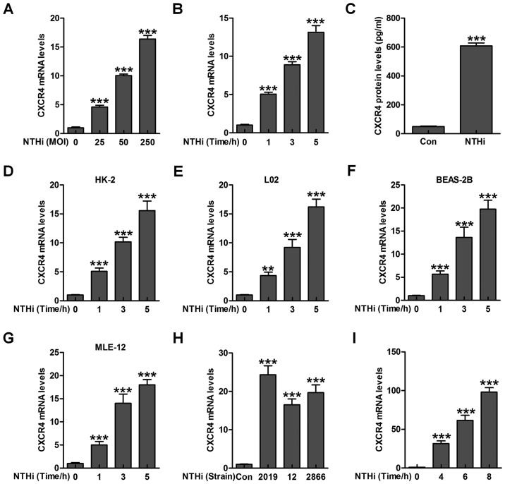
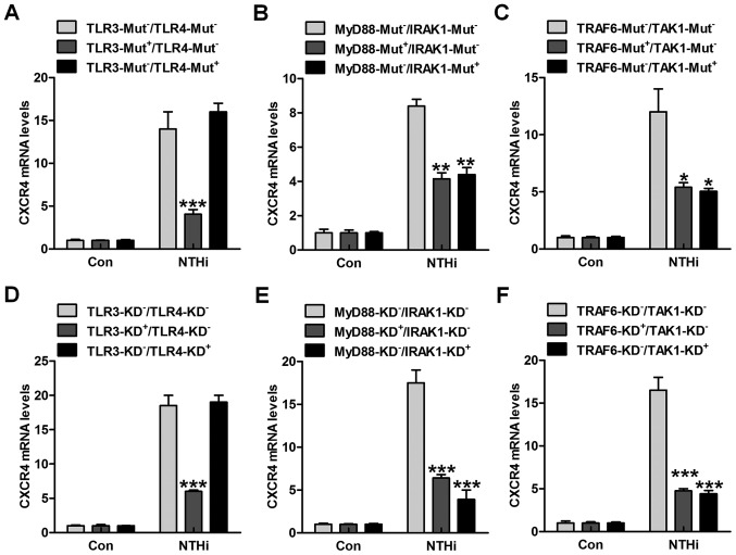
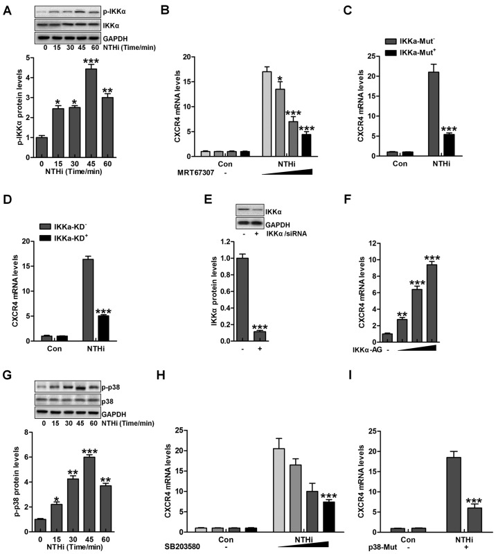
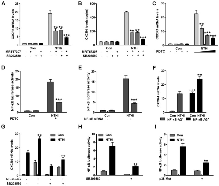
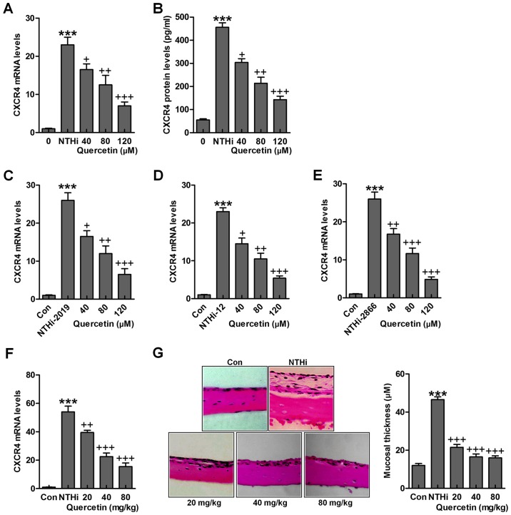
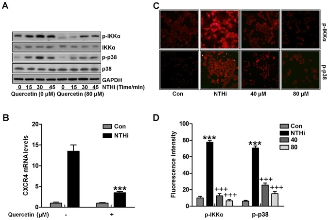
Otitis media is one of the most common bacterial infections in children, contributing to hearing loss. A vital bacterial pathogen leading to otitis media development is the nontypeable Haemophilus influenzae (NTHi). Inflammation response is reported as an important characristic for otitis media. Chemokine CXC receptor 4 (CXCR4) is a 352-amino acid seven-span transmembrane G-protein coupled receptor, essential for inflammatory response. However, the possible molecular mechanism indicating the alteration of CXCR4 modulated by NTHi is poorly known. In the present study, NTHi enhanced CXCR4 expression through phosphorylation of IKKα and p38, which relied on nuclear factor-κB (NF-κB) translocation in vitro as well as in the middle ear of mice in vivo. Previously, quercetin, a natural production mainly isolated from rutin, has shown anti-inflammatory effects. Here, we report that quercetin suppressed NTHi-induced CXCR4 expression levels in vitro and in vivo. Quercetin blocked CXCR4 activation through direct IKKβ phosphorylation inhibition, as well as of p38 MAPK restraining. Hence, identification of quercetin may be a potential therapeutic strategy for treating otitis media induced by NTHi through inflammation suppression.


Figures

Figure 1
Figure 1
Chemokine CXC receptor 4 (CXCR4) is activated by nontypeable Haemophilus influenzae (NTHi) in epithelial cells of middle ear. (A) NTHi was used to treat human middle ear epithelial cells (HMEECs) (25, 50 and 250 for MOI) for 6 h, followed by measurement of CXCR4 gene levels through RT-qPCR analysis. (B) CXCR4 mRNA levels were detected via RT-qPCR analysis in NTHi (MOI of 50)-treated HMEECs cells for different times as indicated. (C) HMEECs were exposed to NTHi for 6 h, and CXCR4 expression from protein levels was evaluated through enzyme-linked immunosorbent assay (ELISA) method. (D) The renal epithelial cells (HK2), (E) liver epithelial L02 cells, and (F) human lung epithelial cells (BEAS-2B), as well as (G) mouse lung epithelial cells (MLE-12) were treated with NTHi for different times, and the CXCR4 mRNA levels were tested through RT-qPCR assays. (H) HMEECs were exposed to various strains of NTHi (2019, 12 and 2866) for 6 h. RT-qPCR analysis was applied to explore CXCR4 gene levels. (I) The experimental animals were inoculated with NTHi for various times as indicated, and CXCR4 mRNA levels were calculated in the the middle ear tissue samples through RT-qPCR assays. The representative data are shown as SEM ***p<0.001 vs. the control group without any treatments.
Figure 2
Figure 2
Nontypeable Haemophilus influenzae (NTHi)-stimulated chemokine CXC receptor 4 (CXCR4) activation relies on TLR3/MyD88 signaling pathway. human middle ear epithelial cells (HMEECs) were transfected with the control, (A) TLR3-, TLR4-, (B) MyD88-, IRAK1-, (C) TRAF6- and TAK1-mutant plasmids. Then, the cells were exposed to NTHi for 6 h. The CXCR4 mRNA levels were then measured through RT-qPCR analysis. HMEECs were transfected with the Control siRNA, (D) TLR3-, TLR4-, (E) MyD88-, IRAK1-, (F) TRAF6- and TAK1-siRNA for knockdown. CXCR4 gene levels in cells were evaluated via RT-qPCR analysis. Representative data are shown as SEM. *p<0.05, **p<0.01 and ***p<0.001 vs. the group in the absence of gene mutants or silence.
Figure 3
Figure 3
Nontypeable Haemophilus influenzae (NTHi)-induced chemokine CXC receptor 4 (CXCR4) activation is related to IKKα and p38 MAPK activity. (A) Human middle ear epithelial cells (HMEECs) were exposed to NTHi for different times ranging from 0 to 60 min. Western blot analysis was used to investigate IKKα phosphorylation. *p<0.05, **p<0.01 and ***p<0.001 vs. the control group. (B) HMEECs were administered to IKKα inhibitor MRT67307 at various concentrations (0, 0.5, 1 and 2 µM) for 2 h, followed by CXCR4 gene level measurement through RT-qPCR analysis. *p<0.05, **p<0.01 and ***p<0.001 vs. the NTHi-treated group in the absence of MRT67307 treatment. (C) HMEECs were transfected with the Control, and IKKα-mutant plasmid. CXCR4 expression from the gene levels was determined by the use of RT-qPCR analysis. ***p<0.001 vs. the NTHi-treated group without IKKα mutation. (D) HMEECs were transfected with the Control, and IKKα-silence for knockdown. CXCR4 mRNA expression levels were detected by RT-qPCR assays. ***p<0.001 vs. the NTHi-treated group without IKKα silence. (E) After IKKα knockdown, western blot analysis was carried out to calculate CXCR4 expression from the protein levels. ***p<0.001 vs. the control group. (F) HMEECs were treated with or without IKK activator at different concentrations (0, 0.5, 1 and 2 µM) for 12 h. Next, CXCR4 mRNA levels were determined by RT-qPCR analysis. **p<0.01 and ***p<0.001 vs. the control group without any treatment. (G) HMEECs were exposed to NTHi for different times (0 to 60 min). Then, western blot analysis was performed to calculate phosphorylated p38 activation. *p<0.05, **p<0.01 and ***p<0.001 vs. the control group. (H) HMEECs were pretreated with p38 inhibitor at various concentrations (0, 0.5, 1 and 2 µM) for 2 h, followed by CXCR4 gene level measurement through RT-qPCR analysis. ***p<0.001 vs. the NTHi-treated group without SB203580 treatment. (I) HMEECs were transfected with the Control, and p38-mutants plasmid. CXCR4 expression levels were calculated by RT-qPCR assays. ***p<0.001 vs. the NTHi-treated group without p38 mutation. Representative data are shown as SEM.
Figure 4
Figure 4
Nuclear factor-κB (NF-κB) phosphorylation is involved in nontypeable Haemophilus influenzae (NTHi)-induced chemokine CXC receptor 4 (CXCR4) expression dependent on IKKα and p38 MAPK activity. (A) Human middle ear epithelial cells (HMEECs) were pretreated with IKKα and p38 MAPK inhibitors, MRT67307 (0.5 µM) and SB203580 (0.5 µM), for 2 h, followed by NTHi exposure for 6 h. Then, the mRNA levels of CXCR4 were calculated by RT-qPCR analysis. (B) HMEECs were pretreated with IKKα and p38 MAPK inhibitors, MRT67307 and SB203580, for 2 h, followed by NTHi exposure for 6 h. Then, the CXCR4 protein levels were calculated via enzyme-linked immunosorbent assay (ELISA) method. *p<0.05, **p<0.01 and ***p<0.001 vs. the NTHi-treated group in the absence of MRT67307 and SB203580 treatment. (C) HMEECs were pretreated with NF-κB inhibitor, PDTC (0.5 µM), for 2 h, followed by NTHi exposure for 6 h. Then, CXCR4 gene levels were calculated by RT-qPCR analysis. (D) HMEECs were first transfected with the vector of NF-κB luciferase. Then, the cells were pretreated with PDTC (0.5 µM) for 2 h, followed by NTHi induction for 6 h. Finally, NF-κB promoter activity was detected through luciferase analysis. *p<0.05, **p<0.01 and ***p<0.001 vs. the NTHi-treated group without PDTC treatment. (E) HMEECs were transfected with siRNA control or siRNA NF-κB. Cells were then stimulated with NTHi for another 6 h, and NF-κB promoter activity was evaluated via luciferase analysis. ***p<0.001 vs. the NTHi-treated group without NF-κB silence. (F) HMEECs were transfected with NF-κB. Cells were then stimulated using NTHi for 6 h, and the CXCR4 mRNA levels were calculated. **p<0.01 vs. the NTHi-treated group without NF-κB transfection; +++p<0.001 vs. the control group without NF-κB transfection. (G) HMEECs were treated with NF-κB transfection or SB203580 alone or in combination, followed by CXCR4 mRNA level measurement. **p<0.01 vs. the NTHi-treated group without any treatment; ++p<0.001 vs. the NTHi-treated group after NF-κB transfection in the absence of SB203580. (H) NF-κB luciferase vector was transfected to HMEECs combined with SB203580, followed by NF-κB promoter activity. **p<0.01 vs. the NTHi-treated group without SB203580. (I) HMEECs were transfected with the control, and p38-mutants plasmid after NF-κB luciferase transfection. Then, the NF-κB promoter activity was evaluated via luciferase analysis. The representative data are shown as SEM.
Figure 5
Figure 5
Quercetin inhibits NTHi-triggered chemokine CXC receptor 4 (CXCR4) activation. Human middle ear epithelial cells (HMEECs) were stimulated with nontypeable Haemophilus influenzae (NTHi) for 6 h, followed by quercetin treatment at different concentrations (40, 80 and 120 µM) for 2 h. Then, (A) CXCR4 mRNA levels (B) and protein levels were evaluated by RT-qPCR analysis and enzyme-linked immunosorbent assay (ELISA) method, respectively. HMEECs were stimulated with NTHi strains (C) 2019, (D) 12 and (E) 2866 for 6 h. Quercetin was administered to cells under different conditions for 2 h. Then, RT-qPCR assay was carried out to investigate CXCR4 mRNA levels. (F) Mice were pretreated with 5×10⁷ CFU NTHi for 6 h, followed by quercetin administration through i.p. at 20, 40 and 80 mg/kg for 2 h. CXCR4 mRNA levels in the dissected middle ear tissue samples were calculated. (G) The representative images of middle ear histophathology in NTHi-treated mice with or without quercetin administration exhibited by H&E staining. The quantification of mucosa thickness is shown. The representative data are shown as SEM. *p<0.05, **p<0.01 and ***p<0.001 vs. the control (Con) group. +p<0.05, ++p<0.01 and +++p<0.001 vs. the NTHi group.
Figure 6
Figure 6
Quercetin inhibits nontypeable Haemophilus influenzae (NTHi)-stimulated chemokine CXC receptor 4 (CXCR4) activation through IKKα and p38 MAPK suppression. (A) HMEECs were stimulated by NTHi for different times, followed by quercetin administration (80 µM) for 2 h. Phosphorylated IKKα and p38 protein levels were calculated by western blot analysis. (B) HMEECs were transfected with the IKKα activator. Cells were then exposed to NTHi for 6 h, and treated by quercetin (80 µM) for 2 h. Finally, CXCR4 mRNA expression levels were measured by RT-qPCR analysis. (C) Cells were stimulated with NTHi for 6 h, and then cultured with quercetin (40 and 80 µM) for 2 h. Next, the p-IKKα and p-p38 fluorescent intensity was calculated by the immunofluorescence (IF) analysis. (D) The quantification of p-IKKα and p-p38 positive cells is shown. The representative data are shown as SEM *p<0.05, **p<0.01 and ***p<0.001 vs. the control (Con) group; +p<0.05, ++p<0.01 and +++p<0.001 vs. the NTHi group.

Similar articles

Cited by

References

    1. Morris LM, DeGagne JM, Kempton JB, Hausman F, Trune DR. Mouse middle ear ion homeostasis channels and intercellular junctions. PLoS One. 2012;7:e39004. doi: 10.1371/journal.pone.0039004. - DOI - PMC - PubMed
    1. Topcuoglu N, Keskin F, Ciftci S, Paltura C, Kulekci M, Ustek D, Kulekci G. Relationship between oral anaerobic bacteria and otitis media with effusion. Int J Med Sci. 2012;9:256–261. doi: 10.7150/ijms.4382. - DOI - PMC - PubMed
    1. Bluestone CD, Klein JO. Physiology, pathophysiology and pathogenesis Otitis Media in Infants and Children. 4th edition. BC Decker; 2007. pp. 41–42.
    1. Post JC. Direct evidence of bacterial biofilms in otitis media. Laryngoscope. 2001;111:2083–2094. doi: 10.1097/00005537-200112000-00001. - DOI - PubMed
    1. Darrow DH, Dash N, Derkay CS. Otitis media: Concepts and controversies. Curr Opin Otolaryngol Head Neck Surg. 2003;11:416–423. doi: 10.1097/00020840-200312000-00002. - DOI - PubMed

MeSH terms