. 2018 Jun;41(6):3211-3220.
doi: 10.3892/ijmm.2018.3566. Epub 2018 Mar 14.

hTERT peptide fragment GV1001 demonstrates radioprotective and antifibrotic effects through suppression of TGF‑β signaling

Affiliations

hTERT peptide fragment GV1001 demonstrates radioprotective and antifibrotic effects through suppression of TGF‑β signaling

Wei Chen et al. Int J Mol Med. 2018 Jun.

Abstract

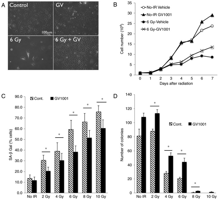
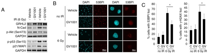
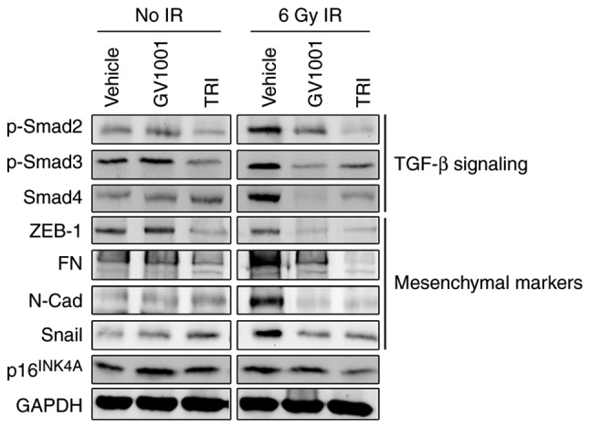
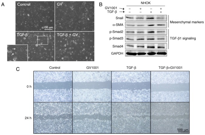
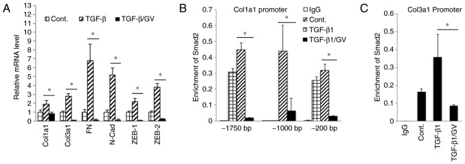
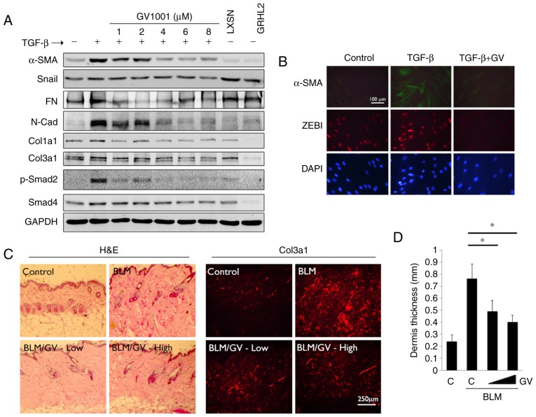
GV1001 is a 16‑amino acid peptide derived from the human telomerase reverse transcriptase (hTERT) protein (616‑626; EARPALLTSRLRFIPK), which lies within the reverse transcriptase domain. Originally developed as an anticancer vaccine, GV1001 demonstrates diverse cellular effects, including anti‑inflammatory, tumor suppressive and antiviral effects. In the present study, the radioprotective and antifibrotic effects of GV1001 were demonstrated through suppressing transforming growth factor‑β (TGF‑β) signaling. Proliferating human keratinocytes underwent premature senescence upon exposure to ionizing radiation (IR), however, treatment of cells with GV1001 allowed the cells to proliferate and showed a reduction in senescent phenotype. GV1001 treatment notably increased the levels of Grainyhead‑like 2 and phosphorylated (p‑)Akt (Ser473), and reduced the activation of p53 and the level of p21/WAF1 in irradiated keratinocytes. It also markedly suppressed the level of TGF‑β signaling molecules, including p‑small mothers against decapentaplegic (Smad)2/3 and Smad4, and TGF‑β target genes, including zinc finger E‑box binding homeobox 1, fibronectin, N‑cadharin and Snail, in irradiated keratinocytes. Furthermore, GV1001 suppressed TGF‑β signaling in primary human fibroblasts and inhibited myofibroblast differentiation. Chromatin immunoprecipitation revealed that GV1001 suppressed the binding of Smad2 on the promoter regions of collagen type III α1 chain (Col3a1) and Col1a1. In a dermal fibrosis model in vivo, GV1001 treatment notably reduced the thickness of fibrotic lesions and the synthesis of Col3a1. These data indicated that GV1001 ameliorated the IR‑induced senescence phenotype and tissue fibrosis by inhibiting TGF‑β signaling and may have therapeutic effects on radiation‑induced tissue damage.


Figures

Figure 1
Figure 1
GV1001 attenuates the IR-induced premature senescence phenotype of NHOKs. (A) Rapidly proliferating NHOKs were exposed to 6 Gy IR and maintained in culture for 10 days in the presence or absence of GV1001 (1 µM) (original magnification, ×100; scale bar, 100 µm; n=3). (B) Cell proliferation kinetics were determined in NHOKs irradiated at 6 Gy IR with GV1001 (1 µM) or vehicle control. (C) NHOKs were exposed to IR at 2–10 Gy in the presence or absence of GV1001 (1 µM) and stained for SA β-Gal. Positively stained cells were counted and plotted. Error bars represent the mean ± standard deviation. (D) NHOKs exposed to 2–10 Gy IR were plated at low density for colony formation in the presence or absence of GV1001 (1 µM). After 10–14 days, the number of colonies were counted and plotted. Error bars represent the mean ± standard deviation. *P<0.05. NHOKs, normal human oral keratinocytes; IR, ionizing radiation; Cont, control; SA β-Gal, senescence-associated β-galactosidase.
Figure 2
Figure 2
GV1001 treatment reduces the level of DNA double strand breaks in NHOKs exposed to IR. (A) Western blot analysis was performed with NHOKs 10 days following exposure to 6 Gy IR with or without GV1001 (1 µM) for GRHL2, N-Cad, p-Akt (Ser473), p-p53 (Ser15) and p21/WAF1. GAPDH was used as a loading control. (B) NHOKs were exposed to 6 Gy IR in the presence or absence of GV1001 (1 µM), were stained for 53BP1 and were viewed under confocal microscopy to detect 53BP1 intranuclear foci. DAPI staining revealed the nuclei (original magnification of left 2 panels, ×100; scale bar, 100 µm; n=3; original magnification of right panels, ×500; scale bar, 20 µm; n=3). (C) Percentages of cells with 53BP1 or γ-H2AX intranuclear foci (>3 foci per nucleus) were counted in ≥10 various fields from each experiment and plotted. Error bars represent the mean ± standard deviation. *P<0.05. NHOKs, normal human oral keratinocytes; IR, ionizing radiation; GRHL2, Grainyhead-like 2; N-Cad, N-Cadherin; p-, phosphorylated.
Figure 3
Figure 3
GV1001 suppresses TGF-β signaling and EMT in NHOKs exposed to IR. NHOKs were exposed to 6 Gy IR and maintained in culture for 10 days in the presence of GV1001 (1 µM) or TRI (1 µM). Western blot analysis was performed for TGF-β signaling molecules, p-Smad2/3 and Smad4, and TGF-β target mesenchymal markers, ZEB1, FN, N-Cad and Snail. GAPDH was used as a loading control. NHOKs, normal human oral keratinocytes; IR, ionizing radiation; TGF-β, transforming growth factor-β; TRI, TGF-β receptor inhibitor; Smad, small mothers against decapentaplegic; p-, phosphorylated; ZEB1, zinc finger E-box binding homeobox 1; FN, fibronectin; N-Cad, N-Cadherin.
Figure 4
Figure 4
GV1001 suppresses TGF-β-induced EMT in NHOKs. (A) Rapidly proliferating NHOKs were exposed to TGF-β (10 ng/ml) for 10 days to induce EMT (shown in islet, arrows). The same cultures were also maintained in the presence or absence of GV1001 (1 µM) (original magnification, ×100; scale bar, 100 µm; n=3). (B) Western blot analysis was performed with NHOKs exposed to TGF-β (10 ng/ml) with GV1001 (1 µM) or the control for Snail, α-SMA, p-Smad2/3 and Smad4. GAPDH was used as a loading control. (C) Cell migration of SCC4 cells exposed to TGF-β (10 ng/ml) with or without GV1001 (1 µM) for 24 h (original magnification, ×40; scale bar, 500 µm; n=3). NHOKs, normal human oral keratinocytes; EMT, epithelial-mesencyhmal transition; TGF-β, transforming growth factor-β; α-SMA, α-smooth muscle actin; Smad, small mothers against decapentaplegic; p-, phosphorylated; GV, GV1001.
Figure 5
Figure 5
GV1001 inhibits Smad2 binding to target gene promoters. (A) Reverse transcription-quantitative polymerase chain reaction analysis was performed on NHOFs exposed to TGF-β (10 ng/ml) with GV1001 (1 µM) or control for 10 days, for the gene expression of Col1a1, Col3a1, FN, N-Cad, Zeb1 and Zeb2. (B and C) NHOFs were cultured with 10 ng/ml TGF-β and GV1001 (1 µM) for 10 days. Chromatin immunoprecipitation was performed for Smad2 enrichment on Col1a1 and Col3a1 promoter regions. Error bars indicate the mean ± standard deviation. *P<0.05. NHOKs, normal human oral keratinocytes; TGF-β, transforming growth factor-β; Col1a1, collagen type I α1 chain; Col3a1, collagen type III α1 chain; ZEB1, zinc finger E-box binding homeobox 1; FN, fibronectin; N-Cad, N-Cadherin; Smad, small mothers against decapentaplegic; Cont, control; GV, GV1001.
Figure 6
Figure 6
GV1001 attenuates the dermal fibrotic lesions induced by BLM. (A) NHOF cultures were treated with 10 ng/ml TGF-β for 10 days and GV1001 was added at varying concentrations between 1 and 8 µM. Western blot analysis was performed with whole cell extracts for α-SMA, Snail, FN, N-Cad, Col1a1 and Col3a1, and p-Smad2 and Smad4. GAPDH was used as a loading control. NHOFs with ectopic overexpression of GRHL2 were also included as a negative control, as GRHL2 inhibited fibrogenic differentiation and TGF-β signaling. (B) IFS was performed for α-SMA and ZEB1 in NHOFs treated with 10 ng/ml TGF-β and GV1001 (1 µM) for 10 days (original magnification, ×100; scale bar, 100 µm; n=3). (C) C57BL/6 mice were exposed to BLM (100 µg/ml) with or without GV1001, administered at low (1 mg/kg) or high (5 mg/kg) doses, by daily subcutaneous injection into the dorsal flank. Following 4 weeks of BLM injection, mice were sacrificed for histological examination by H&E staining. IFS was also performed with the skin biopsy samples for Col3a1 deposition in mice exposed to BLM with or without GV1001 (original magnification, ×100; scale bar, 250 µm; n=5). (D) Quantitation of dermal thickness was plotted in the groups with or without BLM and GV1001 administration. Error bars indicate the mean ± standard deviation. *P<0.05. NHOKs, normal human oral keratinocytes; BLM, bleomycin; IFS, immunofluorescence staining; TGF-β, transforming growth factor-β; α-SMA, α-smooth muscle actin; Col1a1, collagen type I α1 chain; Col3a1, collagen type III α1 chain; ZEB1, zinc finger E-box binding homeobox 1; FN, fibronectin; N-Cad, N-Cadherin; Smad, small mothers against decapentaplegic; p-, phosphorylated; GRHL2, Grainyhead-like 2; H&E, hematoxylin and eosin; GV, GV1001; C, control.

References

    1. Kim HR, Christensen R, Park NH, Sapp P, Kang MK, Park NH. Elevated expression of hTERT is associated with dysplastic cell transformation during human oral carcinogenesis in situ. Clin Cancer Res. 2001;7:3079–3086. - PubMed
    1. Kim NW, Piatyszek MA, Prowse KR, Harley CB, West MD, Ho PL, Coviello GM, Wright WE, Weinrich SL, Shay JW. Specific association of human telomerase activity with immortal cells and cancer. Science. 1994;266:2011–2015. doi: 10.1126/science.7605428. - DOI - PubMed
    1. Greener M. Telomerase: The search for a universal cancer vaccine. Mol Med Today. 2000;6:257. doi: 10.1016/S1357-4310(00)01731-7. - DOI - PubMed
    1. Brunsvig PF, Aamdal S, Gjertsen MK, Kvalheim G, Markowski-Grimsrud CJ, Sve I, Dyrhaug M, Trachsel S, Møller M, Eriksen JA, Gaudernack G. Telomerase peptide vaccination: A phase I/II study in patients with non-small cell lung cancer. Cancer Immunol Immunother. 2006;55:1553–1564. doi: 10.1007/s00262-006-0145-7. - DOI - PMC - PubMed
    1. Bernhardt SL, Gjertsen MK, Trachsel S, Møller M, Eriksen JA, Meo M, Buanes T, Gaudernack G. Telomerase peptide vaccination of patients with non-resectable pancreatic cancer: A dose escalating phase I/II study. Br J Cancer. 2006;95:1474–1482. doi: 10.1038/sj.bjc.6603437. - DOI - PMC - PubMed