

Biphasic activation of nuclear factor-κB and expression of p65 and c-Rel following traumatic neuronal injury
- PMID: 29568960
- PMCID: PMC5881643
- DOI: 10.3892/ijmm.2018.3567
Biphasic activation of nuclear factor-κB and expression of p65 and c-Rel following traumatic neuronal injury
Abstract
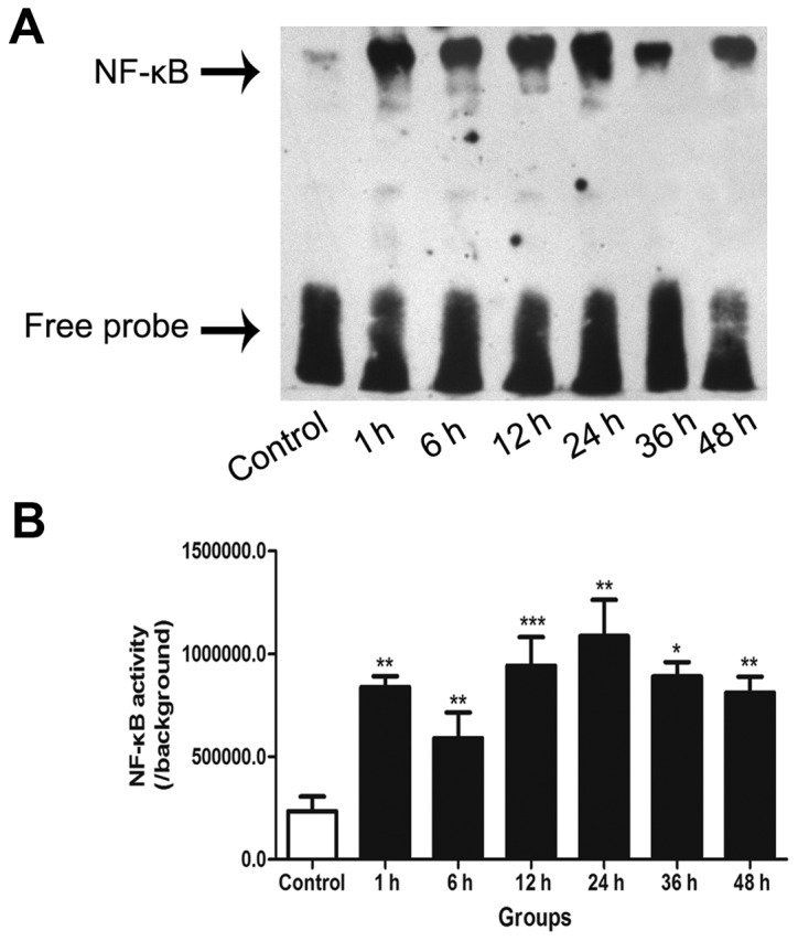
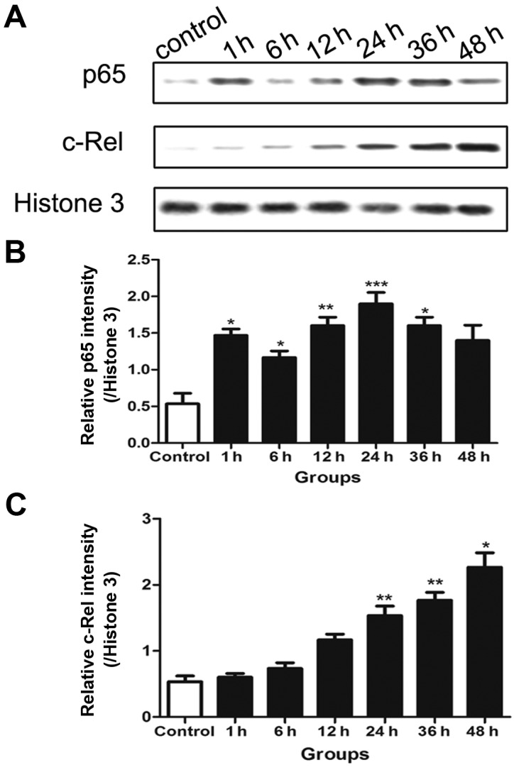
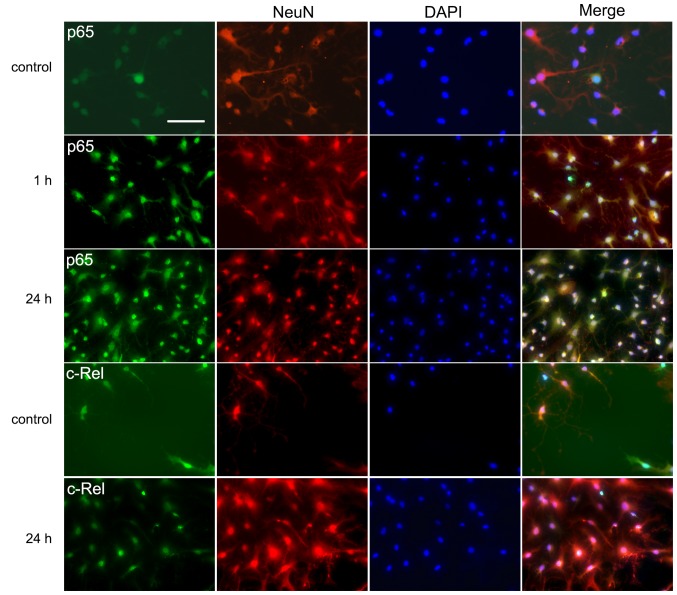
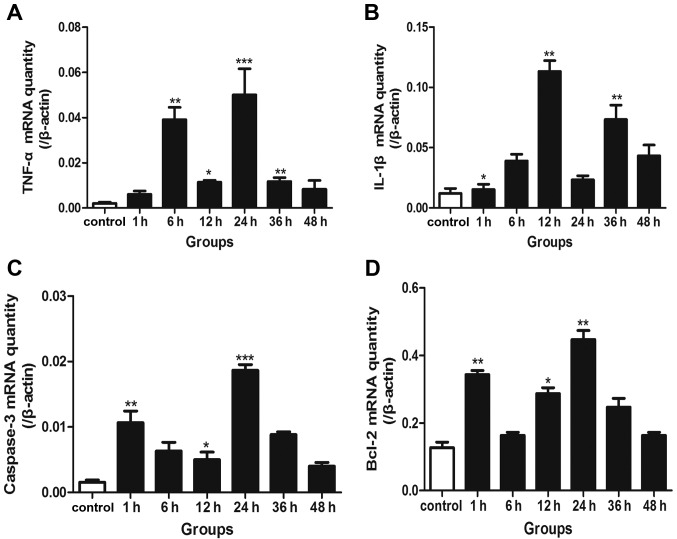
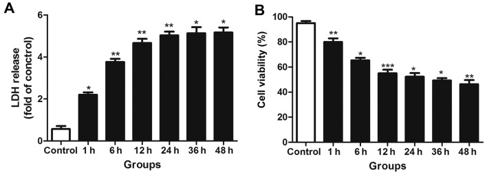
The transcription factor nuclear factor-κB (NF-κB) has been shown to function as a key regulator of cell death or survival in neuronal cells. Previous studies indicate that the biphasic activation of NF-κB occurs following experimental neonatal hypoxia-ischemia and subarachnoid hemorrhage. However, the comprehensive understanding of NF-κB activity following traumatic brain injury (TBI) is incomplete. In the current study, an in vitro model of TBI was designed to investigate the NF-κB activity and expression of p65 and c-Rel subunits following traumatic neuronal injury. Primary cultured neurons were assigned to control and transected groups. NF-κB activity was detected by electrophoretic mobility shift assay. Western blotting and immunofluorescence were used to investigate the expression and distribution of p65 and c-Rel. Reverse transcription-quantitative polymerase chain reaction was performed to assess the downstream genes of NF-κB. Lactate dehydrogenase (LDH) quantification and trypan blue staining were used to estimate the neuronal injury. Double peaks of elevated NF-κB activity were observed at 1 and 24 h following transection. The expression levels of downstream genes exhibited similar changes. The protein levels of p65 also presented double peaks while c-Rel was elevated significantly in the late stage. The results of the trypan blue staining and LDH leakage assays indicated there was no sustained neuronal injury during the late peak of NF-κB activity. In conclusion, biphasic activation of NF-κB is induced following experimental traumatic neuronal injury. The elevation of p65 and c-Rel levels at different time periods suggests that within a single neuron, NF-κB may participate in different pathophysiological processes.
Figures
Similar articles
-
Biphasic activation of nuclear factor kappa B and expression of p65 and c-Rel after traumatic brain injury in rats.Inflamm Res. 2014 Feb;63(2):109-15. doi: 10.1007/s00011-013-0677-1. Epub 2013 Oct 22. Inflamm Res. 2014. PMID: 24146067
-
Participation of the E3-ligase TRIM13 in NF-κB p65 activation and NFAT-dependent activation of c-Rel upon T-cell receptor engagement.Int J Biochem Cell Biol. 2014 Sep;54:217-22. doi: 10.1016/j.biocel.2014.07.012. Epub 2014 Aug 1. Int J Biochem Cell Biol. 2014. PMID: 25088585
-
Distinct nuclear factor-kappaB/Rel proteins have opposing modulatory effects in glutamate-induced cell death in HT22 cells.Neurochem Int. 2005 Dec;47(8):545-55. doi: 10.1016/j.neuint.2005.07.010. Epub 2005 Sep 22. Neurochem Int. 2005. PMID: 16183169
-
Nuclear factor-kappaB/Rel proteins: a point of convergence of signalling pathways relevant in neuronal function and dysfunction.Biochem Pharmacol. 1999 Jan 1;57(1):1-7. doi: 10.1016/s0006-2952(98)00214-7. Biochem Pharmacol. 1999. PMID: 9920279 Review.
-
Rel/NF-kappaB transcription factors: key mediators of B-cell activation.Immunol Rev. 2000 Aug;176:134-40. doi: 10.1034/j.1600-065x.2000.00615.x. Immunol Rev. 2000. PMID: 11043773 Review.
Cited by
-
Long noncoding RNA NKILA transferred by astrocyte-derived extracellular vesicles protects against neuronal injury by upregulating NLRX1 through binding to mir-195 in traumatic brain injury.Aging (Albany NY). 2021 Mar 3;13(6):8127-8145. doi: 10.18632/aging.202618. Epub 2021 Mar 3. Aging (Albany NY). 2021. PMID: 33686956 Free PMC article.
-
Effect of miR-506-3p on Proliferation and Apoptosis of Airway Smooth Muscle Cells in Asthmatic Mice by Regulating CCL2 Gene Expression and Mediating TLR4/NF-κB Signaling Pathway Activation.Mol Biotechnol. 2021 May;63(5):410-423. doi: 10.1007/s12033-021-00309-8. Epub 2021 Feb 27. Mol Biotechnol. 2021. PMID: 33638773
-
Absence of Glia Maturation Factor Protects from Axonal Injury and Motor Behavioral Impairments after Traumatic Brain Injury.Exp Neurobiol. 2020 Jun 30;29(3):230-248. doi: 10.5607/en20017. Exp Neurobiol. 2020. PMID: 32565489 Free PMC article.
-
ITF2357 regulates NF-κB signaling pathway to protect barrier integrity in retinal pigment epithelial cells.FASEB J. 2024 Mar 15;38(5):e23512. doi: 10.1096/fj.202301592R. FASEB J. 2024. PMID: 38430220 Free PMC article.
-
Andrographolide Alleviates Acute Brain Injury in a Rat Model of Traumatic Brain Injury: Possible Involvement of Inflammatory Signaling.Front Neurosci. 2018 Sep 20;12:657. doi: 10.3389/fnins.2018.00657. eCollection 2018. Front Neurosci. 2018. PMID: 30294256 Free PMC article.
References
MeSH terms
Substances
LinkOut - more resources
Full Text Sources
Other Literature Sources
Medical
