Review
. 2018 Jun;23(4):665-685.
doi: 10.1007/s00775-018-1552-x. Epub 2018 Mar 22.

The NMR contribution to protein-protein networking in Fe-S protein maturation

Affiliations
Review

The NMR contribution to protein-protein networking in Fe-S protein maturation

Lucia Banci et al. J Biol Inorg Chem. 2018 Jun.

Erratum in

Abstract

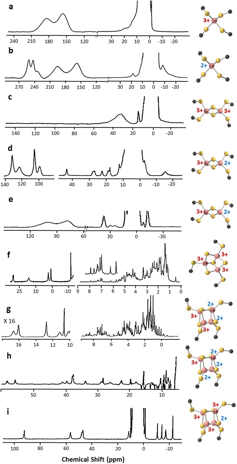
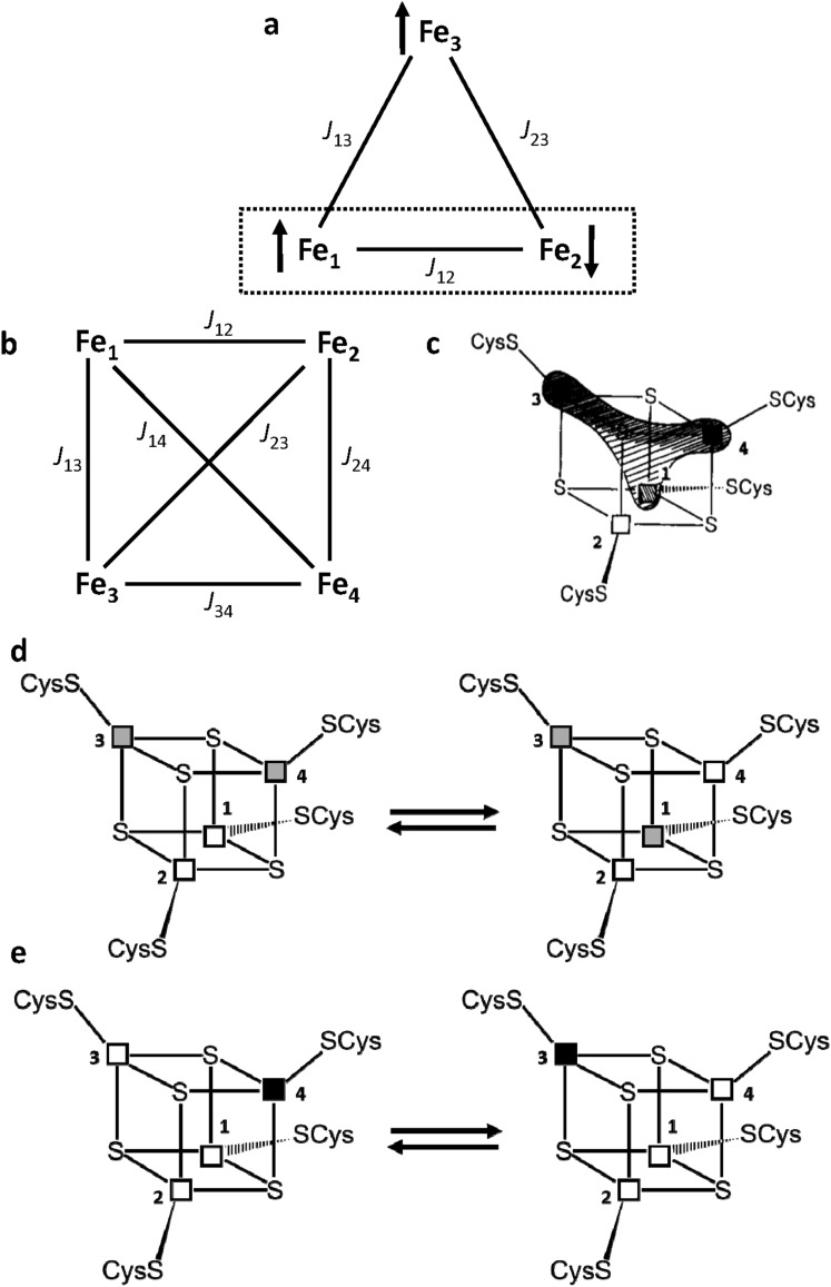
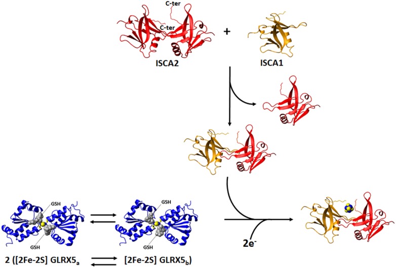
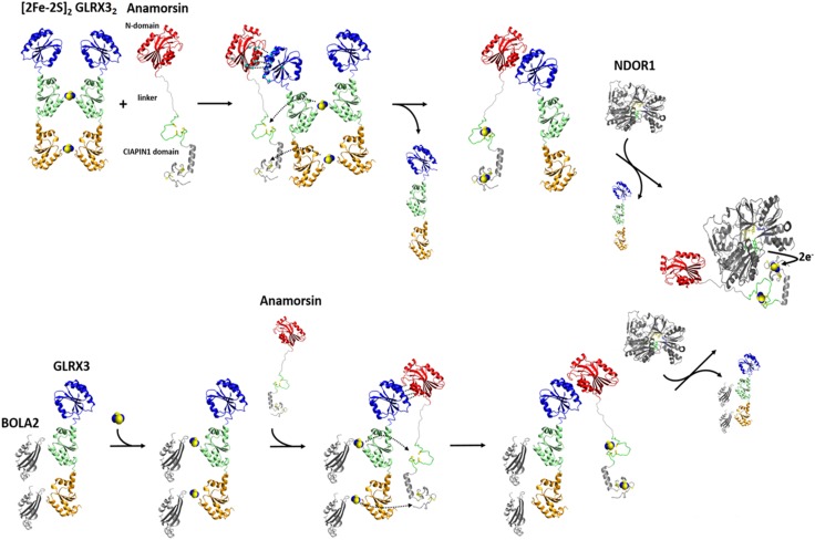
Iron-sulfur proteins were among the first class of metalloproteins that were actively studied using NMR spectroscopy tailored to paramagnetic systems. The hyperfine shifts, their temperature dependencies and the relaxation rates of nuclei of cluster-bound residues are an efficient fingerprint of the nature and the oxidation state of the Fe-S cluster. NMR significantly contributed to the analysis of the magnetic coupling patterns and to the understanding of the electronic structure occurring in [2Fe-2S], [3Fe-4S] and [4Fe-4S] clusters bound to proteins. After the first NMR structure of a paramagnetic protein was obtained for the reduced E. halophila HiPIP I, many NMR structures were determined for several Fe-S proteins in different oxidation states. It was found that differences in chemical shifts, in patterns of unobserved residues, in internal mobility and in thermodynamic stability are suitable data to map subtle changes between the two different oxidation states of the protein. Recently, the interaction networks responsible for maturing human mitochondrial and cytosolic Fe-S proteins have been largely characterized by combining solution NMR standard experiments with those tailored to paramagnetic systems. We show here the contribution of solution NMR in providing a detailed molecular view of "Fe-S interactomics". This contribution was particularly effective when protein-protein interactions are weak and transient, and thus difficult to be characterized at high resolution with other methodologies.

Keywords: CIA machinery; Fe–S proteins; Hyperfine interactions; ISC machinery; Interactomics; NMR spectroscopy.


Figures

Fig. 1
Fig. 1
1D 1H NMR spectra of different Fe–S cluster types. 400 MHz 1D 1H NMR spectra of Fe3+ (a) and Fe2+ (b) C. pasteurianum rubredoxin, acquired at 308 K (adapted from [13]); c 200 MHz 1D 1H NMR spectrum of [2Fe–2S]2+ P. umbilicalis ferredoxin, acquired at 303 K [15]; d 360 MHz 1D 1H NMR spectrum of [2Fe–2S]+ P. umbilicalis ferredoxin, recorded at 303 K [25]; e 400 MHz 1D 1H NMR spectrum of [2Fe–2S]+ human ferredoxin, acquired at 303 K [201]; f 500 MHz 1D 1H NMR spectrum of [3Fe–4S]+ P. furiosus ferredoxin, recorded at 303 K [33]; 600 MHz 1D 1H NMR spectra of [4Fe–4S]2+, g [40] and [4Fe–4S]3+, i [60] E. halophila HIPIP II, recorded at 300 K; h 600 MHz 1D 1H NMR spectrum of [4Fe–4S]+ C. acidi urici ferredoxin, acquired at 298 K [43]
Fig. 2
Fig. 2
Magnetic coupling and electronic distribution in [3Fe–4S] and [4Fe–4S] clusters. Schematic representation of the spin frustration in a [3Fe–4S] cluster (a) and of the coupling scheme in a [4Fe–4S] cluster (b). ce Electronic distribution in the [4Fe–4S]3+ clusters of HiPIPs: c the extra electron can be unevenly distributed among the iron ions Fe1, Fe3 and Fe4; d a chemical equilibrium between two different electronic distributions in the cluster, where mixed-valence Fe2.5+–Fe2.5+ iron ion pairs are represented as grey squares and purely Fe3+–Fe3+ pairs are represented as white squares; e illustration of the resonance between two limit formulas. Fe3+ and Fe2+ ions are represented as white and black squares, respectively
Fig. 3
Fig. 3
The 1H–15N IR-HSQC-AP NMR experiment: a new tool for paramagnetic Fe–S proteins. Schematic drawings of the pulse sequence of the 1H–15N IR-HSQC-AP NMR experiment. The inversion recovery delay τ and the coherence transfer delay δ must be chosen according to, respectively, T1 and T2 relaxation properties of the signals of interest. b Efficiency of an INEPT transfer function at different 1H T2 values: b 100 ms, c 10 ms, d 5 ms, e 2 ms, f 1 ms, g 0.5 ms. Relaxation is neglected in a. Letters have been drawn at the correspondence of the maximum values for each transfer function. A dashed line is shown at the 2.65 ms of INEPT step (94 Hz for 1H–15N J coupling). A solid line is shown in correspondence of 10% transfer efficiency. The latter is a limit threshold below which direct excitation of 15N spins should replace the INEPT step in the first part of the sequence. c Optimized 1H–15N IR-HSQC-AP experiment vs. d standard 1H–15N HSQC experiment acquired on 500 MHz at 298 K on the [2Fe–2S]2+-CIAPIN1 domain of human anamorsin
Fig. 4
Fig. 4
Solution NMR as a tool to investigate weak, transient protein–protein interactions in Fe–S protein maturation pathways. Weak and transient protein–protein interactions are detected by following backbone NH chemical shift changes occurring in a standard 1H–15N HSQC NMR experiment and in a 1H–15N IR-HSQC-AP NMR experiment, upon titrating 15N-labeled protein with the unlabeled protein partner and vice versa. Standard 1H–15N HSQC experiments allow the identification of protein–protein interacting regions far from the paramagnetic Fe–S cluster (showed in cyano), and to estimate the dissociation constant (Kd) of the observed interaction. 1H–15N IR-HSQC-AP NMR experiment allows to identify protein–protein interacting regions close to the paramagnetic Fe–S cluster (showed in yellow). 1D 1H NMR experiment provides information on the kind of Fe–S cluster(s) bound or assembled on a target protein or protein–protein complex, on the redox state(s) of the cluster(s), and on the cluster ligands
Fig. 5
Fig. 5
The NMR contribution to the investigation of [4Fe–4S] clusters formation in the mitochondrial iron–sulfur cluster assembly machinery. On the basis of standard and paramagnetic systems-tailored NMR experiments, we provide a model for the transfer of two [2Fe–2S]2+ clusters from GLRX5 to ISCA1–ISCA2 hetero-dimeric complex: in solution dimeric [2Fe–2S]2+ GLRX5 has two states in equilibrium with each other, differing in the binding mode of the GSH molecules ([2Fe–2S] GLRX5a and [2Fe–2S] GLRX5b); dimeric [2Fe–2S]2+ GLRX5 specifically transfers the cluster to apo ISCA1–ISCA2 hetero-dimeric complex via an associative process that involves a transient protein–protein intermediate; [2Fe–2S] GLRX5b is more reactive than [2Fe–2S] GLRX5a to donate the cluster; ISCA1–ISCA2 hetero-dimeric complex is obtained in solution by exchanging one subunit of the ISCA2 symmetric dimer with one subunit of ISCA1, which, as isolated protein, is present in solution in a monomer–dimer equilibrium; the two [2Fe–2S]2+ clusters received preferentially by [2Fe–2S] GLRX5b are reductively coupled on ISCA1–ISCA2 hetero-dimeric complex to form a [4Fe–4S]2+ cluster
Fig. 6
Fig. 6
The NMR contribution to the elucidation of  the GLRX3-dependent anamorsin maturation pathway. Standard 1H–15N-HSQC and 1H–15N IR-HSQC-AP NMR experiments, combined with UV–Vis and EPR spectroscopy, showed that GLRX3 forms a 1:1 hetero-dimeric complex with anamorsin, in which both clusters from [2Fe–2S]2 GLRX32 are transferred to anamorsin. In its mature holo state anamorsin interacts with NDOR1, forming a specific protein complex, where the anamorsin unstructured linker tightly interact with NDOR1, while the C-terminal CIAPIN1 domain of anamorsin, containing the [2Fe–2S] redox center, only transiently interacts, through complementary charged residues, with the FMN-binding domain of NDOR1 to perform the electron transfer reaction. Standard NMR experiments showed that the [2Fe–2S]2 GLRX3–BOLA22 hetero-complex transfers in vitro both [2Fe–2S]2+ clusters to apo anamorsin, producing its mature holo state, and that this process goes via the same protein–protein recognition mechanism operating in the GLRX3-anamorsin interaction, i.e., specifically occurring between the N-terminal domains of the two proteins. The BOLA2–GLRX3 complex might be released in solution upon the interaction of holo anamorsin with NDOR1

References

    1. Kowalsky A. Biochemistry. 1965;4:2382–2388.
    1. Wüthrich K. Proc Natl Acad Sci USA. 1969;63:1071–1078. - PMC - PubMed
    1. McDonald CC, Phillips WD, Vinogradov SN. Biochem Biophys Res Commun. 1969;36:442–449. - PubMed
    1. Phillips WD, Poe M, Weiher JF, McDonald CC, Lovenberg W. Nature. 1970;227:574–577. - PubMed
    1. Poe M, Phillips WD, McDonald CC, Lovenberg W. Proc Natl Acad Sci USA. 1970;65:797–804. - PMC - PubMed

Publication types

MeSH terms

Substances

LinkOut - more resources