. 2018 Jun;16(6):1198-1210.
doi: 10.1111/jth.14008. Epub 2018 May 8.

Novel assay demonstrates that coronary artery disease patients have heightened procoagulant platelet response

Affiliations

Novel assay demonstrates that coronary artery disease patients have heightened procoagulant platelet response

L Pasalic et al. J Thromb Haemost. 2018 Jun.

Abstract

Essentials Procoagulant platelets can be detected using GSAO in human whole blood. Stable coronary artery disease is associated with a heightened procoagulant platelet response. Agonist-induced procoagulant platelet response is not inhibited by aspirin alone. Collagen plus thrombin induced procoagulant platelet response is partially resistant to clopidogrel.

Summary: Background Procoagulant platelets are a subset of highly activated platelets with a critical role in thrombin generation. Evaluation of their clinical utility in thrombotic disorders, such as coronary artery disease (CAD), has been thwarted by the lack of a sensitive and specific whole blood assay. Objectives We developed a novel assay, utilizing the cell death marker, GSAO [(4-(N-(S-glutathionylacetyl)amino)phenylarsonous acid], and the platelet activation marker, P-selectin, to identify procoagulant platelets in whole blood by flow cytometry. Patients/Methods Using this assay, we characterized the procoagulant platelet population in healthy controls and a cohort of patients undergoing elective coronary angiography. Results In patients with CAD, compared with patients without CAD, there was a heightened procoagulant platelet response to thrombin (25.2% vs. 12.2%), adenosine diphosphate (ADP) (7.8% vs. 2.7%) and thrombin plus collagen (27.2% vs. 18.3%). The heightened procoagulant platelet potential in CAD patients was not associated with other markers of platelet function, including aggregation, dense granule release and activation of α2b β3 integrin. Although dual antiplatelet therapy (DAPT) was associated with partial suppression of procoagulant platelets, this inhibitory effect on a patient level could not be predicted by aggregation response to ADP and was not fully suppressed by clopidogrel. Conclusions We report for the first time that procoagulant platelets can be efficiently detected in a few microliters of whole blood using the cell death marker, GSAO, and the platelet activation marker, P-selectin. A heightened procoagulant platelet response may provide insight into the thrombotic risk of CAD and help identify a novel target for antiplatelet therapies in CAD.

Keywords: coronary artery disease; flow cytometry; platelet activation; platelet aggregation inhibitors; platelet function studies; procoagulant platelets.


Conflict of interest statement

L. Pasalic, V. M. Chen and P. J. Hogg have patent PCT/AU2015/000638 pending. The other authors state that they have no conflict of interest.

Figures

Figure 1
Figure 1
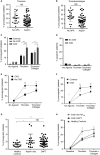
Gating strategy for detection of procoagulant platelets in human whole blood using the cytoplasmic cell death marker (GSAO). (A) Platelets are defined as CD41a positive but CD45 negative events. (B) Threshold values for P‐selectin and GSAO‐positive regions are set using a thrombin plus collagen‐stimulated sample stained with IgG isotype control and control GSCA, respectively. GSAO+/P‐selectin+ (Procoag) quadrant (outlined in red) contains procoagulant platelets. GSAO+/P‐selectin− (Apopt) quadrant (outlined in green) contains apoptotic platelets. (C) Very few platelets show the procoagulant phenotype (P‐selectin+/GSAO+) in unstimulated whole blood. (D) Following stimulation with thrombin plus collagen a significant proportion of platelets exhibited the procoagulant phenotype. (E) Characterization of the stimulus‐induced procoagulant platelet response in human whole blood. (F) Synergistic interaction between thrombin and collagen occurred at all levels of individual agonist concentrations. Normalized agonist dose (maximal [100%] for collagen [100 μg mL−1] and thrombin [6.3 U mL−1]) expressed as percent maximal is shown on a logarithmic scale. A representative example is shown. (G) Apoptotic platelets are a very small subpopulation that does not increase with agonist stimulation. (E, G) Mean ± SEM, n = 16–35. One‐way anova with Sidak's correction for multiple comparisons, *P < 0.05, **P < 0.0001. ADP, adenosine diphosphate; GSAO, [(4‐(N‐(S‐glutathionylacetyl)amino)phenylarsonous acid]; PLA, platelet leukocyte aggregates; WBC, white blood cells. [Color figure can be viewed at wileyonlinelibrary.com]
Figure 2
Figure 2
GSAO‐based assay has advantages over other markers of cell death for identification of procoagulant platelets in human whole blood. (A) Whole blood is the preferred substrate for demonstration of procoagulant platelet potential. Citrated blood samples from individuals were prepared as whole blood (WB), platelet‐rich plasma (PRP) or washed platelet (WP) preparations. Each preparation was treated with thrombin (2 U mL−1), collagen (10 μg mL−1), or a combination of both, then stained with GSAO/P‐selectin. WB demonstrated higher levels of GSAO+/P‐selectin+ platelets compared with PRP or WP from the same draw. (B) Most GSAO+ platelets externalized PS (lactadherin+); however, a significant proportion of lactadherin+ platelets did not take up GSAO. Pseudocolors are used to indicate population density. (C) Thrombin (2 U mL−1)‐treated WB samples were stained either with a standard panel of markers or a modified panel where GSAO was replaced with the amine reactive dye Zombie NIR, fixed and analyzed by flow cytometry. Procoagulant platelets identified by GSAO were not identified by Zombie NIR (n = 3, paired‐t‐test, *P < 0.05). GSAO, [(4‐(N‐(S‐glutathionylacetyl)amino)phenylarsonous acid]. [Color figure can be viewed at wileyonlinelibrary.com]
Figure 3
Figure 3
CAD is associated with a heightened procoagulant platelet potential and loss of synergism between thrombin and collagen that is not affected by aspirin. DAPT ameliorates thrombin hypersensitivity in CAD but only partially suppresses thrombin and collagen. (A, B) Blood samples (1–4 per individual) were collected during coronary angiography from 60 patients on either aspirin or no antiplatelet agents. Procoagulant platelets were assayed in whole blood samples treated with thrombin (2 U mL−1) or thrombin (2 U mL−1) plus collagen (10 μg mL−1). Data are shown as the percentage of platelets with the procoagulant phenotype (GSAO+/ P‐selectin+). Linear mixed effects models, two‐tailed tests, *P < 0.05. (C) Platelets from patients with CAD showed increased stimulus‐induced procoagulant potential compared to those without CAD and healthy controls. Two‐way anova analysis with Tukey correction for multiple comparisons. Mean ± SEM, n = 11 (CAD), n = 16 (no CAD) and n = 10 (healthy controls). P < 0.0001. (D) Apoptotic platelet subset (GSAO+/P‐selectin−) did not vary according to the presence of CAD. Two‐way anova analysis with Tukey correction for multiple comparisons. Mean ± SEM, n = 11 (CAD), n = 17 (no CAD). (E) Platelets from CAD patients showed increased procoagulant response to thrombin and thrombin collagen compared with angiographically normal patients (**P < 0.01). However, CAD patients failed to respond to further stimulation with addition of collagen to thrombin (thrombin vs. thrombin plus collagen, paired t‐test, P = 0.9684). (F) Validation of heightened procoagulant platelet response to thrombin and thrombin collagen in an independent cohort of CAD patients not treated with P2Y12 antagonists. Mean ± SEM, n = 11 (CAD) and n = 10 (healthy controls), t‐test **P < 0.01. (G) Without stimulation, circulating procoagulant platelets were elevated in CAD patients compared with healthy controls (t‐test *P < 0.05, **P < 0.01)). Levels of circulating procoagulant platelets were not statistically different between patients on aspirin or DAPT. (H) DAPT reduced the hypersensitivity to thrombin and restored the synergism between thrombin and collagen stimulation compared with thrombin alone (t test **P < 0.01) in the validation CAD cohort. DAPT (n = 22), no P2Y12 (n = 11). APA, antiplatelet agents; CAD, coronary artery disease; DAPT, dual antiplatelet therapy; GSAO, [(4‐(N‐(S‐glutathionylacetyl)amino)phenylarsonous acid]; NS, not significant.
Figure 4
Figure 4
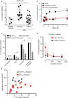
Clopidogrel abrogates the abnormal response to ADP and thrombin but not to a thrombin and collagen combination in patients with CAD, and shows a discordant inhibitory effect on platelet aggregation and procoagulant potential. (A) CAD patients without P2Y12 exposure show an increased procoagulant response to ADP 5 μm (2.7% ± 1.8% vs. 7.6% ± 3.2%, *P < 0.05) that is abrogated in patients on DAPT (7.6% ± 3.2% vs. 4.1% ± 2.5%, t‐test **P < 0.01). (B) Platelets from patients with CAD showed heightened procoagulant response to ADP (≥ 0.625 μm) compared with healthy controls. Treatment with clopidogrel restored the pattern of response. n = 3, t‐test *P < 0.05, **P < 0.01. (C) Compared with patients without P2Y12 antagonists, patients on clopidogrel showed significant inhibition of procoagulant platelet formation in response to ADP 5 μm (*P < 0.05) or thrombin 2 U mL−1 (*P < 0.05), but not thrombin 2 U mL−1 and collagen 10 μg mL−1 (t‐test P = 0.99). Patients on prasugrel or ticagrelor demonstrated significant inhibition of thrombin/collagen‐induced procoagulant platelets compared with patients on clopidogrel (27.2 ± 4.5% vs. 12.0 ± 1.8%, n = 8–22, t‐test P < 0.05); however, there was no additional inhibition of procoagulant response to ADP (P = 0.3) or thrombin (P = 0.13) in patients on prasugrel or ticagrelor vs. clopidogrel. (D) Ex‐vivo spiking of ticagrelor into subjects’ blood stimulated with thrombin (2 U mL−1) and collagen (10 mg mL−1) showed that ticagrelor reduced procoagulant platelet formation in CAD patients on aspirin only (n = 5), to the same level as in healthy controls (n = 6). (E) The GSAO‐based assay allows identification of clopidogrel‐treated patients with heightened procoagulant platelet response despite adequate suppression of aggregation. Correlation between ADP‐induced platelet aggregation responses (y‐axis) measured in whole blood and the procoagulant platelet response to thrombin plus collagen (x‐axis). Dotted horizontal green lines represent the published normal range for ADP‐induced aggregation in healthy blood donors 43; dotted vertical black lines represent the 10th and 90th percentile range for thrombin plus collagen‐induced procoagulant platelets in healthy controls. Treatment with P2Y12 antagonists is associated with suppression of ADP‐induced aggregation; however, a significant proportion of procoagulant responses remain above the 90th percentile. ADP, adenosine diphosphate; AUC, area under curve; CAD, coronary artery disease; DAPT, dual antiplatelet therapy; GSAO, [(4‐(N‐(S‐glutathionylacetyl)amino)phenylarsonous acid]. [Color figure can be viewed at wileyonlinelibrary.com]

Similar articles

Cited by

References

    1. Heemskerk JWM, Mattheij NJA, Cosemans JMEM. Platelet‐based coagulation: different populations, different functions. J Thromb Haemost 2013; 11: 2–16. - PubMed
    1. McFadyen JD, Jackson SP. Differentiating haemostasis from thrombosis for therapeutic benefit. Thromb Haemost 2013; 110: 859–67. - PubMed
    1. Bouchard BA, Silveira JR, Tracy PB. Chapter 21 ‐ interactions between platelets and the coagulation system In: Michelson AD, ed. Platelets (3rd Edn). Cambridge: Academic Press, 2013: 425–51.
    1. Munnix ICA, Kuijpers MJE, Auger J, Thomassen CMLGD, Panizzi P, van Zandvoort MAM, Rosing J, Bock PE, Watson SP, Heemskerk JWM. Segregation of platelet aggregatory and procoagulant microdomains in thrombus formation: regulation by transient integrin activation. Arterioscler Thromb Vasc Biol 2007; 27: 2484–90. - PMC - PubMed
    1. Libby P, Theroux P. Pathophysiology of coronary artery disease. Circulation 2005; 111: 3481–8. - PubMed

Publication types

MeSH terms