

Resolvin D1 attenuates liver ischaemia/reperfusion injury through modulating thioredoxin 2-mediated mitochondrial quality control
- PMID: 29569721
- PMCID: PMC5980610
- DOI: 10.1111/bph.14212
Resolvin D1 attenuates liver ischaemia/reperfusion injury through modulating thioredoxin 2-mediated mitochondrial quality control
Abstract
Background and purpose: Liver ischaemia and reperfusion (IR) injury is a sterile inflammatory response involving production of ROS. Mitochondrial homeostasis is maintained by mitochondrial quality control (QC). Thioredoxin (TRX) 2 is a key mitochondrial redox-sensitive protein. Resolvin D1 (RvD1), a specialized pro-resolving lipid mediator, exerts anti-inflammatory and antioxidant activities. We investigated mechanisms of RvD1 protection against IR-induced oxidative damage to the liver, focusing on TRX2-mediated mitochondrial QC.
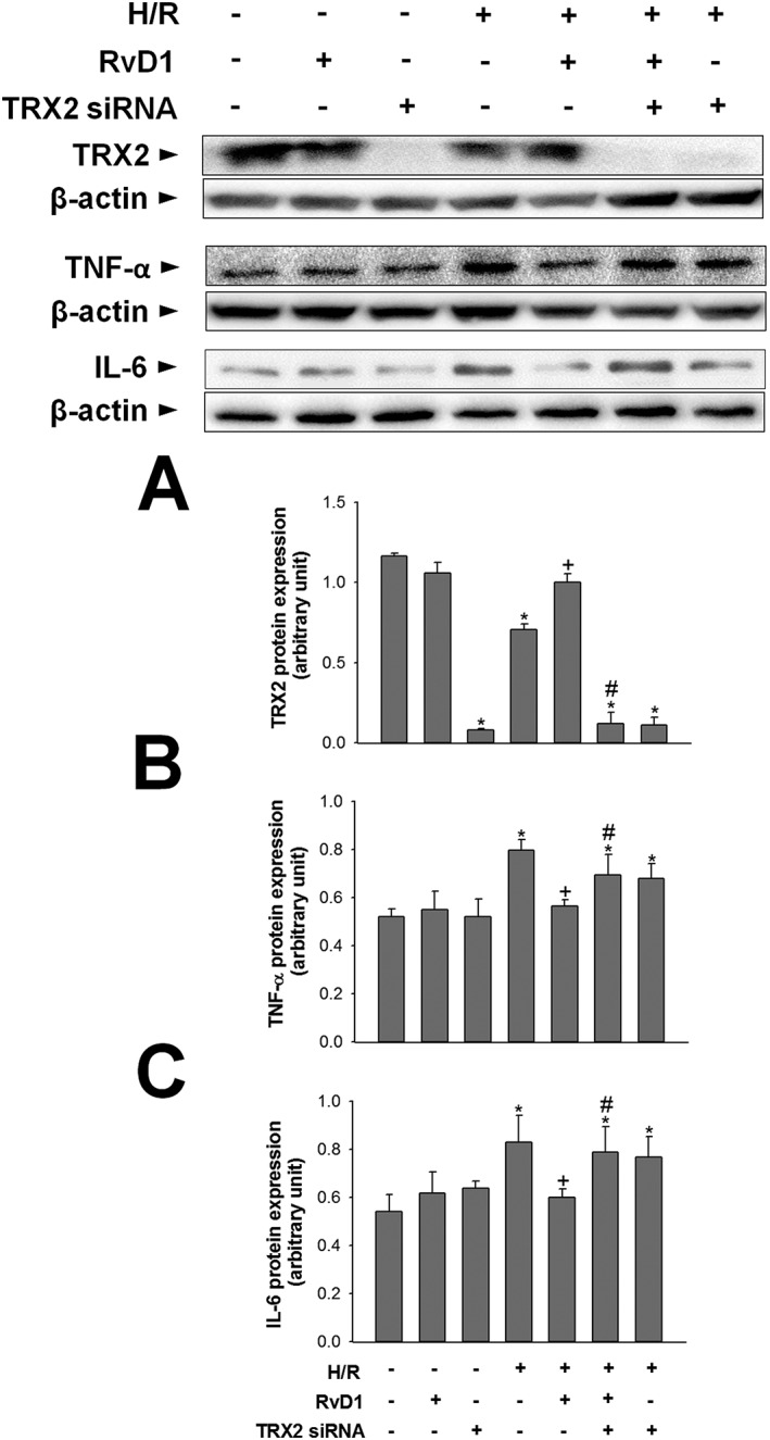
Experimental approach: Mice underwent partial warm IR. RvD1 was administered 1 h before ischaemia and immediately prior to reperfusion. Human liver carcinoma HepG2 cells were exposed to hypoxia/reoxygenation and transfected with TRX2 siRNA. Immunohistochemistry, Western blotting and enzyme assays were used to follow changes in mitochondrial structure and function.
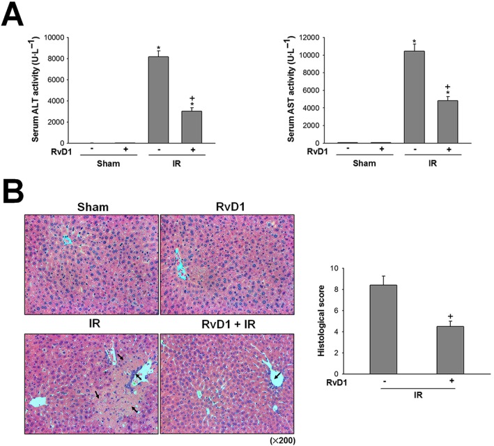
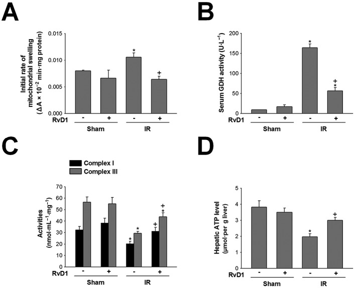
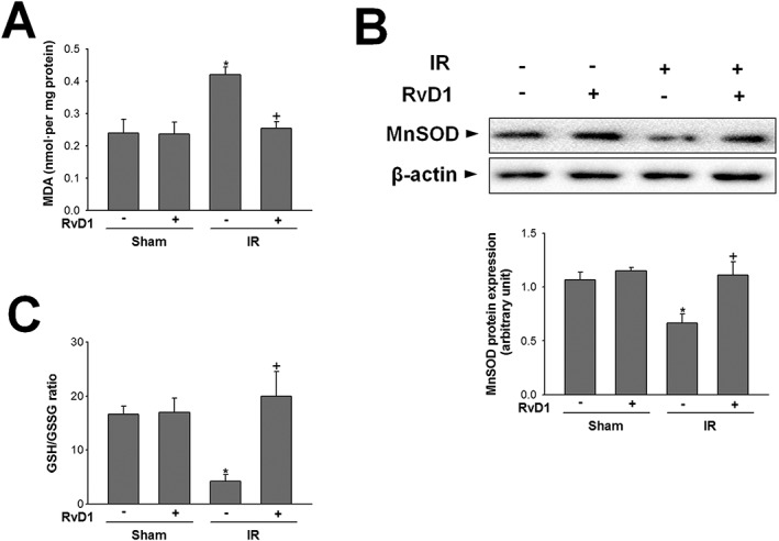
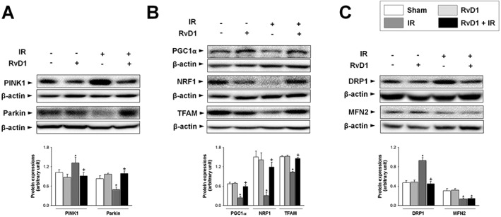
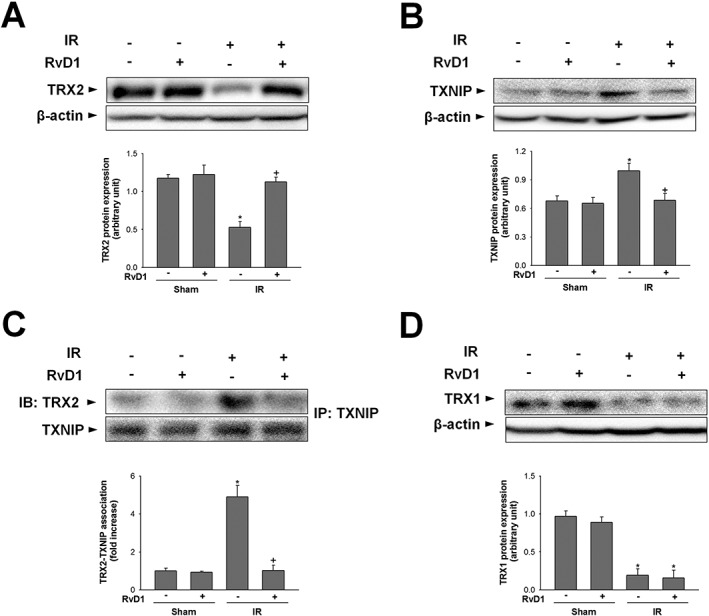
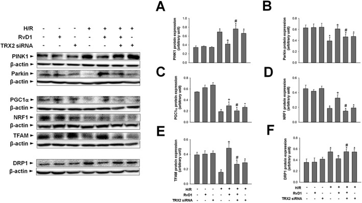
Key results: RvD1 attenuated hepatocellular damage following IR, assessed by serum aminotransferase activities and histology. RvD1 reduced mitochondrial swelling, lipid peroxidation and glutamate dehydrogenase release. Impaired activities of mitochondrial complexes I and III were restored by RvD1. RvD1 enhanced expression of the mitophagy-related protein, Parkin and inhibited accumulation of PTEN-induced putative kinase 1. RvD1 restored levels of mitochondrial biogenesis proteins including PPARγ coactivator 1α, nuclear respiratory factor 1 and mitochondrial transcription factor A and mtDNA level. RvD1 attenuated the increase in levels of the mitochondrial fission-related protein, dynamin-related protein 1. IR reduced TRX2 levels while increasing TRX2 association with TRX-interacting protein. RvD1 attenuated these changes. The regulatory effects of RvD1 on mitochondrial QC were abolished by TRX2 knockdown.
Conclusions and implications: We suggest that RvD1 ameliorated IR-induced hepatocellular damage by regulating TRX2-mediated mitochondrial QC.
© 2018 The British Pharmacological Society.
Figures
Similar articles
-
Resolvin D1 activates the sphingosine-1-phosphate signaling pathway in murine livers with ischemia/reperfusion injury.Biochem Biophys Res Commun. 2019 Jul 5;514(4):1058-1065. doi: 10.1016/j.bbrc.2019.05.041. Epub 2019 May 13. Biochem Biophys Res Commun. 2019. PMID: 31097221
-
Genipin protects the liver from ischemia/reperfusion injury by modulating mitochondrial quality control.Toxicol Appl Pharmacol. 2017 Aug 1;328:25-33. doi: 10.1016/j.taap.2017.05.002. Epub 2017 May 3. Toxicol Appl Pharmacol. 2017. PMID: 28477916
-
Resolvin D1 protects the liver from ischemia/reperfusion injury by enhancing M2 macrophage polarization and efferocytosis.Biochim Biophys Acta. 2016 Sep;1861(9 Pt A):1025-1035. doi: 10.1016/j.bbalip.2016.06.002. Epub 2016 Jun 15. Biochim Biophys Acta. 2016. PMID: 27317426
-
Resolvin D1 protects against hepatic ischemia/reperfusion injury in rats.Int Immunopharmacol. 2015 Sep;28(1):322-7. doi: 10.1016/j.intimp.2015.06.017. Epub 2015 Jun 25. Int Immunopharmacol. 2015. PMID: 26118631
-
The role of Resolvin D1 in liver diseases.Prostaglandins Other Lipid Mediat. 2022 Jun;160:106634. doi: 10.1016/j.prostaglandins.2022.106634. Epub 2022 Mar 12. Prostaglandins Other Lipid Mediat. 2022. PMID: 35292355 Review.
Cited by
-
Noncoding RNA Roles in Pharmacogenomic Responses to Aspirin: New Molecular Mechanisms for an Old Drug.Biomed Res Int. 2021 Dec 9;2021:6830560. doi: 10.1155/2021/6830560. eCollection 2021. Biomed Res Int. 2021. PMID: 34926688 Free PMC article. Review.
-
Mitochondrial quality control in hepatic ischemia-reperfusion injury.Heliyon. 2023 Jun 27;9(7):e17702. doi: 10.1016/j.heliyon.2023.e17702. eCollection 2023 Jul. Heliyon. 2023. PMID: 37539120 Free PMC article. Review.
-
Resolvin D1 attenuated liver injury caused by chronic ethanol and acute LPS challenge in mice.FASEB J. 2023 Jan;37(1):e22705. doi: 10.1096/fj.202200778R. FASEB J. 2023. PMID: 36520060 Free PMC article.
-
Preservation of Mitochondrial Health in Liver Ischemia/Reperfusion Injury.Biomedicines. 2023 Mar 20;11(3):948. doi: 10.3390/biomedicines11030948. Biomedicines. 2023. PMID: 36979927 Free PMC article. Review.
-
Liver metabolomics reveals potential mechanism of Jieduan-Niwan formula against acute-on-chronic liver failure (ACLF) by improving mitochondrial damage and TCA cycle.Chin Med. 2023 Nov 30;18(1):157. doi: 10.1186/s13020-023-00858-x. Chin Med. 2023. PMID: 38037150 Free PMC article.
References
-
- Bannenberg GL, Chiang N, Ariel A, Arita M, Tjonahen E, Gotlinger KH et al (2005). Molecular circuits of resolution: formation and actions of resolvins and protectins. J Immunol 174: 4345–4355. - PubMed
Publication types
MeSH terms
Substances
LinkOut - more resources
Full Text Sources
Other Literature Sources
Medical
Research Materials
