. 2018 Sep;70(9):1489-1499.
doi: 10.1002/art.40505. Epub 2018 Jul 25.

An Autoimmune Basis for Raynaud's Phenomenon: Murine Model and Human Disease

Affiliations

An Autoimmune Basis for Raynaud's Phenomenon: Murine Model and Human Disease

D P Ascherman et al. Arthritis Rheumatol. 2018 Sep.

Abstract

Objective: Raynaud's phenomenon (RP) is common in anti-RNP-positive patients with rheumatic diseases but is not itself known to be caused by autoimmunity. The aim of this study was to assess autoantibodies that could mediate this process.

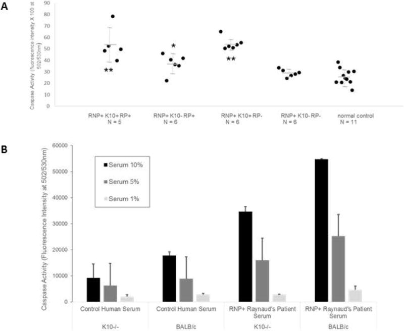
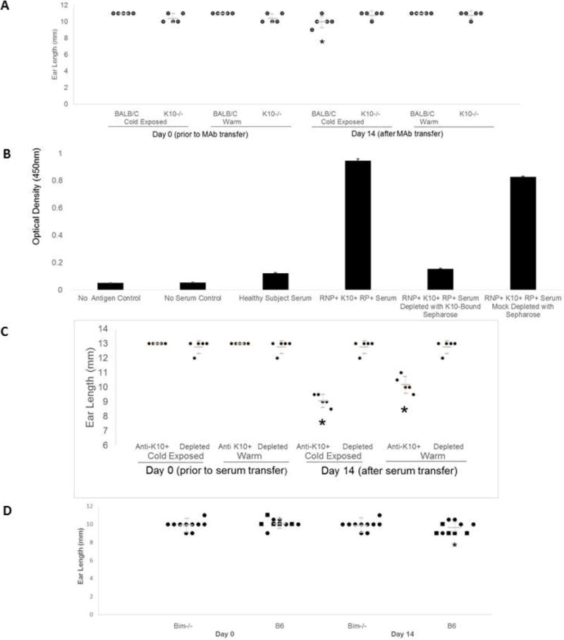
Methods: Antibodies derived from patient sera and from murine models of anti-RNP autoimmunity were screened for the ability to induce RP-like tissue ischemia and endothelial cell apoptosis in murine models and in vitro systems.

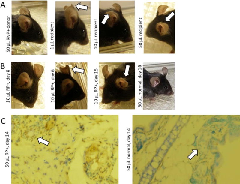
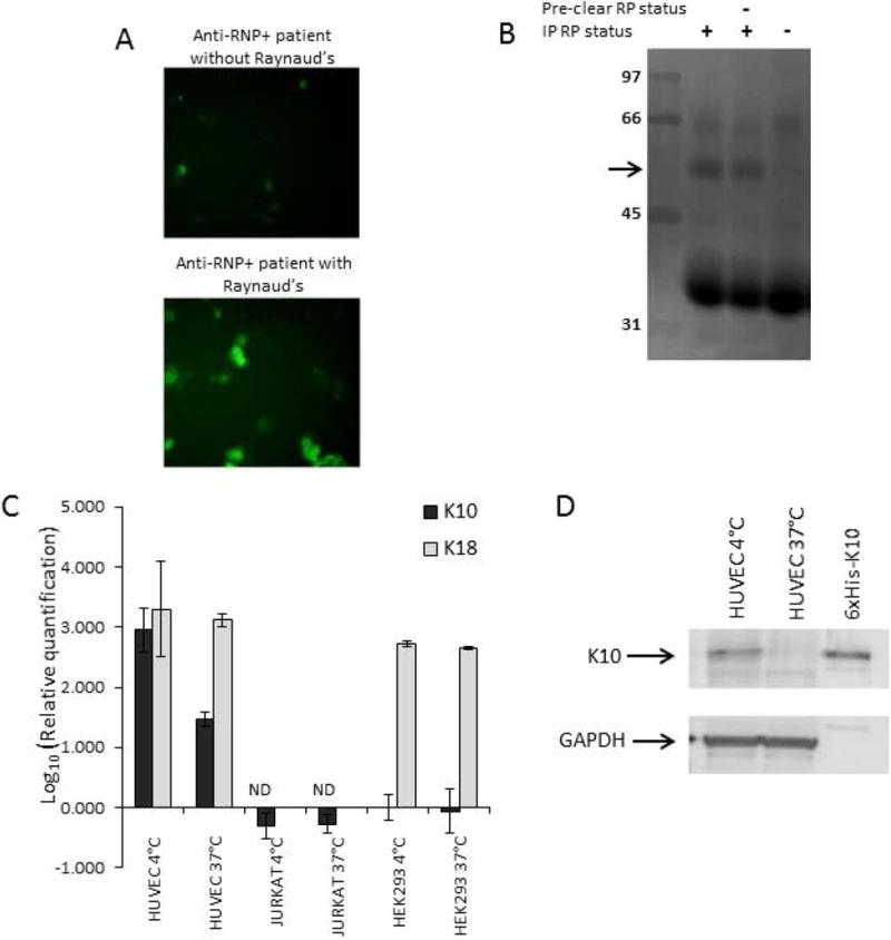
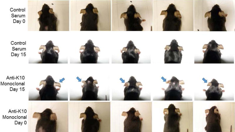
Results: RNP-positive sera from RP patients and murine sera from RNP-positive B cell adoptive transfer recipients induced RP-like tissue ischemia and endothelial cell apoptosis. Proteomic analysis identified cytokeratin 10 (K10) as a candidate autoantigen in RP. Monoclonal anti-K10 antibodies reproduced patterns of ischemic tissue loss and endothelial cell apoptosis; K10 knockout or depletion of anti-K10 activity in serum was protective. Cold exposure enhanced K10 expression and in vivo tissue loss.

Conclusion: Anti-K10 antibodies are sufficient to mediate RP-like ischemia in murine models and are implicated in the pathogenesis of RP in patients with anti-RNP autoimmunity.


Conflict of interest statement

The authors do not have any financial interests that could be perceived as being a conflict of interest. No patents pertaining to the results presented in the paper have been awarded or filed.

Figures

Figure 1
Figure 1
B cell transfer-induced murine model of ischemic Raynaud’s. Progressive ear and tail loss are noted in syngeneic B cell recipients from 70k-immunized HLA-DR4tg B6 mice. In the top three rows, the same mice are shown 7 and 14 days after cell transfer; arrows indicate areas of tissue loss. At the bottom, an additional recipient mouse that lost its tail 14 days after transfer is shown along with the ischemic tail.
Figure 2
Figure 2
Serum transfer of ischemia phenotype. A. Murine serum transfer from ischemic B cell recipients as in Figure 1 induces dose-dependent tissue loss by 14 days (arrows), not seen with B cell donor sera. B. Human Raynaud’s patient serum transfer (10 microliters) following the same protocol as in A induces progressive ear tissue loss (arrows) from Day 0 to Day 6 to Day 15 in the same mouse, not seen with healthy control human serum. C. Raynaud’s serum transfer induces vascular apoptosis. Mice received human Raynaud’s or healthy control sera. Two weeks later, ear tissue (10×) showed increased TUNEL staining (brown) in the Raynaud’s serum recipient compared to the normal control, prominently near the lumen of blood vessels (arrows).
Figure 3
Figure 3
K10 as a cold-inducible target of Raynaud’s-associated antibodies. Cultured cells were kept at 37°C in complete medium (see Methods). Where indicated for cold exposure, cells were exposed to 4°C medium for 1 minute 24 hours before cell harvesting, then returned to 37°C. A. Anti-endothelial activity by immunofluorescence of HUVEC (see Methods) with an RNP+ serum from a Raynaud’s patient. B. Reverse immunophenotyping immunoprecipitation (see Methods) of HUVEC lysates with an anti-RNP+ human Raynaud’s Phenomenon serum identified a ~56kD band (arrow) that was not pre-cleared by an RNP+ serum from a patient without Raynaud’s Phenomenon (RP-), for MSMS analysis. C. Expression and cold-induced upregulation of mRNA levels by real time PCR were quantitated for K10 and control cytokeratin 18 (K18), normalized to expression of 18S ribosomal RNA. The endothelial cell line HUVEC expressed much higher levels of K10 message than epithelial HEK293 cells or lymphoid Jurkat cells at 37°C, and further dramatically upregulated K10 but not K18 expression levels after cold exposure. D. Expression and upregulation of K10 protein in HUVEC. While low levels of K10 protein were present in HUVEC cells at 37°C, the quantity of protein dramatically increased in cold-exposed cells (compared to GAPDH loading control).
Figure 4
Figure 4
Anti-K10 monoclonal antibodies induce ischemic tissue loss. Female 10 week old C57BL/6 mice received 50 μg of anti-mouse anti-K10 monoclonal antibody by IV tail vein infusion (“Anti-K10 Monoclonal”, n = 5), or an equal amount of normal mouse immunoglobulin (“Control Serum”, n = 5). Representative images are shown at Days 0 and 15 for each of the 10 mice. None of the Control Serum mice had any clinical manifestations (0/5). In contrast, 5/5 Anti-Mouse K10 recipients (100%, Fisher’s Exact p = 0.008) developed cyanosis and ischemia of the ears, with progressive tissue loss (bilateral, though most prominent at arrows). Results are representative of 3 separate experiments.
Figure 5
Figure 5
K10-dependent endothelial apoptosis. A. HUVEC were exposed for 24 hours to 10% dilutions of sera from healthy subjects (Ctrl) or RNP+ patients with or without Raynaud’s (RP+ or -) and anti-K10 IgG antibodies (ELISA > 2 S.D. above healthy subject mean to be anti-K10+), loaded with CellEvent Caspase 3/7 Green Detection Reagent (10μM/ml) and assayed for green absorbance in 24 hours in duplicate wells. Anti-K10+ sera induced high levels of caspase activity (an indicator of apoptosis) from patients with or without RP. ** t test p < 0.041 versus K10-RP+ sera, p < 0.003 versus K10-RP- sera, and p <= 0.0001 versus Ctrl K10- sera; * t test p < 0.048 versus K10-RP- sera and p < 0.009 versus Ctrl K10- sera. Results were representative of 3 separate experiments. B. Endothelial cell cultures concurrently generated from BALB/c, and K10-knockout BALB/c mice (see Methods) were exposed to dilutions of K10- control serum or K10+ RNP+ RP+ serum for 24 hours, and assayed for caspase activity as above (representative of 2 separate experiments). The Raynaud’s serum induced increased dose-dependent caspase activity in both K10-intact and K10−/− cells compared to control serum, but much more so in K10-intact cells.
Figure 6
Figure 6
K10−/−, Anti-K10 depletion, or Bim−/− prevent ischemia. 24h before antibody injections, one ear of each mouse was exposed to 4°C for one minute. Ear lengths were measured at baseline and Day 14. A. Anti-K10 MAb (50 μg) injected via tail vein (n = 5). In K10−/−, no ear loss was observed, regardless of cold exposure. BALB/C mice had ear loss after cold exposure in 4/5 mice (*change in ear length versus K10−/− mice T Test p = 0.008). B. Anti-K10+ human Raynaud’s serum depletion by incubation with his6-K10-loaded nickel-Sepharose beads yielded control serum levels of anti-K10 ELISA reactivity. Nickel-Sepharose mock depletion had minimal effect. C. In B6 mice, ear tissue was lost in 5/5 recipients of mock-depleted serum, each worse on the cold-exposed side, but in 0/5 recipients of anti-K10 depleted serum (*Fisher’s Exact p = 0.008). D. Bim−/− and germline control B6 mice (n = 10) received 50 μl anti-K10+ human serum; only cold-exposed ears shown. Squares: ear loss observed; Circles: no ear loss. Bim−/− mice were protected from ear loss versus Bim-intact mice (*Fisher’s Exact p = 0.033).

References

    1. Voulgari PV, Alamanos Y, Papazisi D, Christou K, Papanikolaou C, Drosos AA. Prevalence of Raynaud’s phenomenon in a healthy Greek population. Ann Rheum Dis. 2000;59:206–10. - PMC - PubMed
    1. Greidinger EL, Casciola-Rosen L, Morris SM, Hoffman RW, Rosen A. Autoantibody recognition of distinctly modified forms of the UI-70-kd antigen is associated with different clinical disease manifestations. Arthritis Rheum. 2000;43:881–888. - PubMed
    1. Amanzi L, Braschi F, Fiori G, Galluccio F, Miniati I, Guiducci S, et al. Digital ulcers in scleroderma: staging, characteristics and sub-setting through observation of 1614 digital lesions. Rheumatology (Oxford) 2010;49:1374–82. - PubMed
    1. Chung L, Shapiro L, Fiorentino D, Baron M, Shanahan J, Sule S, et al. MQX-503, a novel formulation of nitroglycerin, improves the severity of Raynaud’s phenomenon: a randomized, controlled trial. Arthritis Rheum. 2009;60:870–7. - PubMed
    1. Cooke JP, Marshall JM. Mechanisms of Raynaud’s disease. Vasc Med. 2005;10:293–307. - PubMed

Publication types