

Seasonal expressions of androgen receptor, estrogen receptors and cytochrome P450 aromatase in the uteri of the wild Daurian ground squirrels (Spermophilus dauricus)
- PMID: 29569876
- PMCID: PMC5820527
- DOI: 10.4081/ejh.2018.2889
Seasonal expressions of androgen receptor, estrogen receptors and cytochrome P450 aromatase in the uteri of the wild Daurian ground squirrels (Spermophilus dauricus)
Abstract
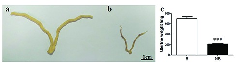
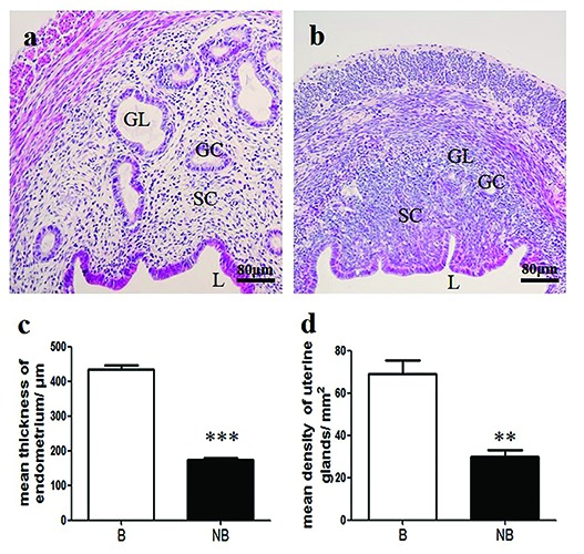
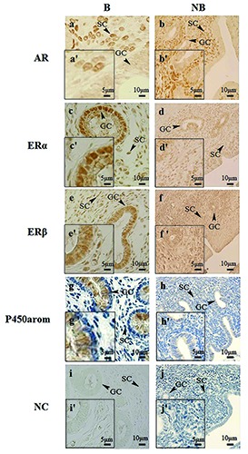
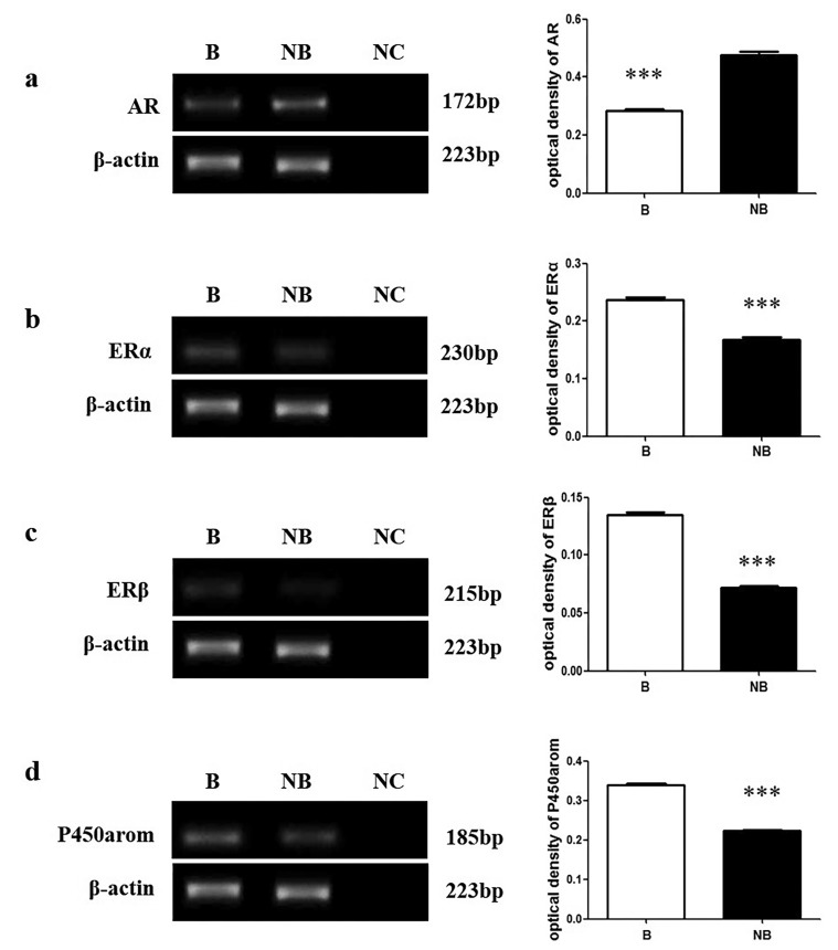
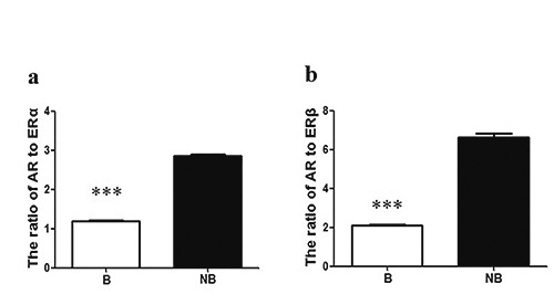
The reproductive tissues including the uterus undergo dramatic changes in seasonal breeders from the breeding to non-breeding seasons. Classically, sex steroid hormones play important roles in the uterine morphology and functions. To clarify the relationship between sex steroid hormones and seasonal changes in the uterine morphology and functions, the wild Daurian ground squirrels (Spermophilus dauricus) were used as seasonal breeder model. And the immunolocalizations and expression levels of androgen receptor (AR), estrogen receptors α and β (ERα and ERβ) and cytochrome P450 aromatase (P450arom) were investigated in the uteri of the wild Daurian ground squirrels in the breeding (April) and the non-breeding (June) seasons via immunohistochemistry, Western blot and RT-PCR. Histologically, the uterine weight, the thickness of endometrium and the glandular density were significantly higher in the uteri of the breeding season than those of the non-breeding season. In both seasons, the immunostaining of AR was only presented in stromal cells of the uteri; the positive staining of ERα and ERβ were localized in stromal cells and glandular cells; P450arom was merely immunolocalized in glandular cells. The protein and mRNA expression levels of ERα, ERβ and P450arom were higher in the uteri of the breeding season than those of the non-breeding season; conversely, the expressions of AR were higher in the uteri of the non-breeding season comparing with those of the breeding season in both protein and mRNA levels. The AR: ER ratio in the uteri of the non-breeding season exceeded the AR: ER ratio in the uteri of the breeding season in the wild Daurian ground squirrels. These results suggested that seasonal changes in the expression levels of AR, ERs and P450arom might be correlated with the uterine morphology and histology changes, and estrogen may play an important autocrine/paracrine role in regulating the uterine functions of the wild Daurian ground squirrels.
Keywords: Androgen receptor; P450arom; estrogen receptor α; estrogen receptor β; uteri; wild Daurian ground squirrel..
Conflict of interest statement
Conflict of interest: The authors declare that they have no competing interests.
Figures

Similar articles
-
Seasonal changes of androgen receptor, estrogen receptors and aromatase expression in the hippocampus of the wild male ground squirrels (Citellus dauricus Brandt).Gen Comp Endocrinol. 2017 Aug 1;249:93-100. doi: 10.1016/j.ygcen.2017.05.009. Epub 2017 May 11. Gen Comp Endocrinol. 2017. PMID: 28502742
-
Seasonal expressions of COX-1, COX-2 and EP4 in the uteri of the wild Daurian ground squirrels (Spermophilus dauricus).Prostaglandins Other Lipid Mediat. 2019 Aug;143:106343. doi: 10.1016/j.prostaglandins.2019.106343. Epub 2019 Jun 10. Prostaglandins Other Lipid Mediat. 2019. PMID: 31195125
-
Seasonal changes of androgen receptor, estrogen receptors and aromatase expression in the medial preoptic area of the wild male ground squirrels (Citellus dauricus Brandt).Eur J Histochem. 2016 May 2;60(2):2621. doi: 10.4081/ejh.2016.2621. Eur J Histochem. 2016. PMID: 27349316 Free PMC article.
-
Aromatase expression in male germ cells.J Steroid Biochem Mol Biol. 2001 Dec;79(1-5):203-8. doi: 10.1016/s0960-0760(01)00137-6. J Steroid Biochem Mol Biol. 2001. PMID: 11850226 Review.
-
Estrogen and androgen receptors: regulators of fuel homeostasis and emerging targets for diabetes and obesity.Trends Endocrinol Metab. 2011 Jan;22(1):24-33. doi: 10.1016/j.tem.2010.10.002. Epub 2010 Nov 5. Trends Endocrinol Metab. 2011. PMID: 21109497 Free PMC article. Review.
Cited by
-
Seasonal expressions of GPR41 and GPR43 in the colon of the wild ground squirrels (<em>Spermophilus dauricus</em>).Eur J Histochem. 2022 Jan 21;66(1):3351. doi: 10.4081/ejh.2022.3351. Eur J Histochem. 2022. PMID: 35057584 Free PMC article.
-
Seasonal patterns of prolactin, prolactin receptor, and STAT5 expression in the ovaries of wild ground squirrels (<em>Citellus dauricus</em> Brandt).Eur J Histochem. 2023 Oct 2;67(4):3825. doi: 10.4081/ejh.2023.3825. Eur J Histochem. 2023. PMID: 37781865 Free PMC article.
-
Seasonal Changes in the Distinct Taxonomy and Function of the Gut Microbiota in the Wild Ground Squirrel (Spermophilus dauricus).Animals (Basel). 2021 Sep 13;11(9):2685. doi: 10.3390/ani11092685. Animals (Basel). 2021. PMID: 34573650 Free PMC article.
-
Twenty years of histochemistry in the third millennium, browsing the scientific literature.Eur J Histochem. 2020 Dec 29;64(4):3213. doi: 10.4081/ejh.2020.3213. Eur J Histochem. 2020. PMID: 33478199 Free PMC article.
-
Seasonal expressions of prolactin, prolactin receptor and STAT5 in the scented glands of the male muskrats (Ondatra zibethicus).Eur J Histochem. 2019 Jan 17;63(1):2991. doi: 10.4081/ejh.2019.2991. Eur J Histochem. 2019. PMID: 30652434 Free PMC article.
References
-
- Maruyama T. Stem cells in the uterus: past, present and future. Semin Reprod Med 2015;33:315-6. - PubMed
-
- Sizemore RJ, Hurst PR, McLeod BJ. Effect of steroid hormones on tissue remodelling and progesterone receptors in the uterus of seasonally anoestrous brushtail possums (Trichosurus vulpecula). Reproduction 2004;127:255-64. - PubMed
-
- Choi JP, Zheng Y, Skulte KA, Handelsman DJ, Simanainen U. Development and characterization of uterine glandular epithelium specific androgen receptor knockout mouse model. Biol Reprod 2015;93:120-34. - PubMed
-
- Fujimoto J, Nishigaki M, Hori M, Ichigo S, Itoh T, Tamaya T. Biological implications of estrogen and androgen effects on androgen receptor and its mRNA levels in human uterine endometrium. Gynecological Endocri - nol 1995;9:149-55. - PubMed
MeSH terms
Substances
LinkOut - more resources
Full Text Sources
Other Literature Sources
Research Materials
Miscellaneous
