

Energy Dispersive X-ray (EDX) microanalysis: A powerful tool in biomedical research and diagnosis
- PMID: 29569878
- PMCID: PMC5907194
- DOI: 10.4081/ejh.2018.2841
Energy Dispersive X-ray (EDX) microanalysis: A powerful tool in biomedical research and diagnosis
Abstract
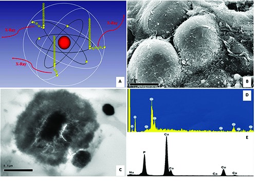
The Energy Dispersive X-ray (EDX) microanalysis is a technique of elemental analysis associated to electron microscopy based on the generation of characteristic Xrays that reveals the presence of elements present in the specimens. The EDX microanalysis is used in different biomedical fields by many researchers and clinicians. Nevertheless, most of the scientific community is not fully aware of its possible applications. The spectrum of EDX microanalysis contains both semi-qualitative and semi-quantitative information. EDX technique is made useful in the study of drugs, such as in the study of drugs delivery in which the EDX is an important tool to detect nanoparticles (generally, used to improve the therapeutic performance of some chemotherapeutic agents). EDX is also used in the study of environmental pollution and in the characterization of mineral bioaccumulated in the tissues. In conclusion, the EDX can be considered as a useful tool in all works that require element determination, endogenous or exogenous, in the tissue, cell or any other sample.
Keywords: Energy Dispersive X-ray (EDX) microanalysis; asbestos; calcifications; nanoparticles.; transmission electron microscopy, scanning electron microscopy.
Figures
References
-
- Scimeca M, Feola M, Romano L, Rao C, Gasbarra E, Bonanno E, et al. Heavy metals accumulation affects bone microarchitecture in osteoporotic patients. Environ Toxicol 2017; 32:1333-42. - PubMed
-
- Barba T, Wach J, Lustig S, Laurent F, Devouassoux-Shisheboran M, Valour F, et al. Metallosis-associated prosthetic joint infection. Med Mal Infect 2015;45:484-7. - PubMed
MeSH terms
Substances
LinkOut - more resources
Full Text Sources
Other Literature Sources
