

Interfacing Cells with Vertical Nanoscale Devices: Applications and Characterization
- PMID: 29570360
- PMCID: PMC6530470
- DOI: 10.1146/annurev-anchem-061417-125705
Interfacing Cells with Vertical Nanoscale Devices: Applications and Characterization
Abstract
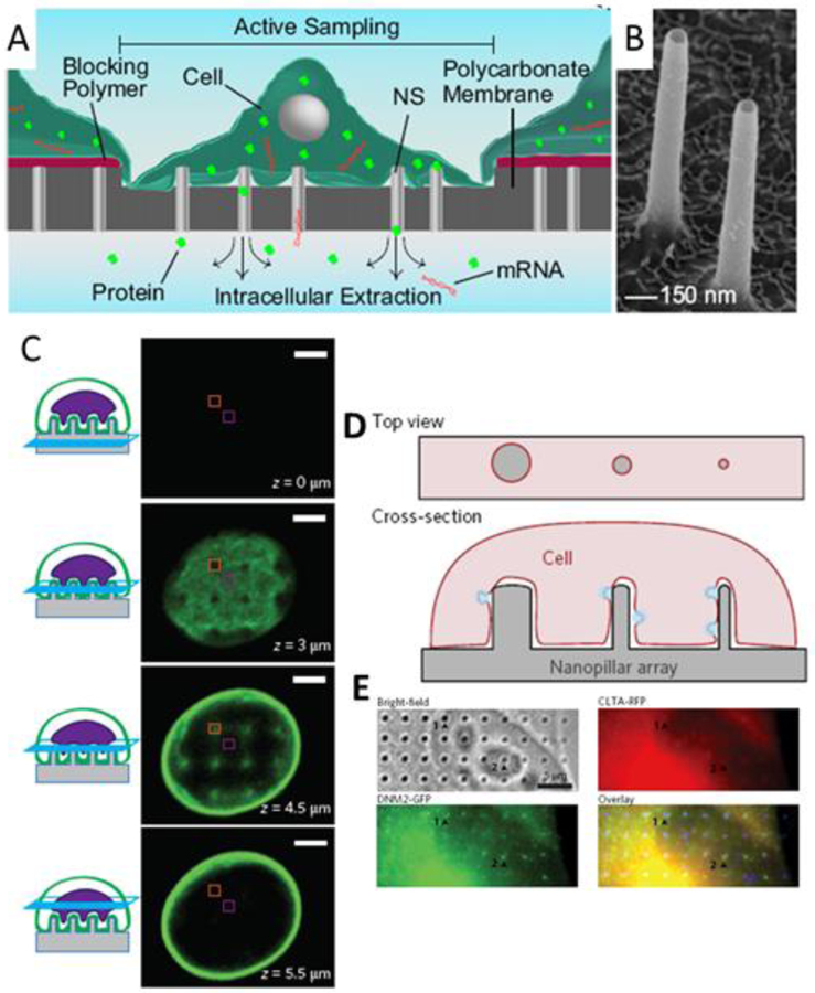
Measurements of the intracellular state of mammalian cells often require probes or molecules to breach the tightly regulated cell membrane. Mammalian cells have been shown to grow well on vertical nanoscale structures in vitro, going out of their way to reach and tightly wrap the structures. A great deal of research has taken advantage of this interaction to bring probes close to the interface or deliver molecules with increased efficiency or ease. In turn, techniques have been developed to characterize this interface. Here, we endeavor to survey this research with an emphasis on the interface as driven by cellular mechanisms.
Keywords: biointerface; electron microscopy; electrophysiology; molecular delivery; vertical structures.
Figures
Similar articles
-
DNA nanostructures interacting with lipid bilayer membranes.Acc Chem Res. 2014 Jun 17;47(6):1807-15. doi: 10.1021/ar500051r. Epub 2014 May 14. Acc Chem Res. 2014. PMID: 24828105
-
Cells Adhering to 3D Vertical Nanostructures: Cell Membrane Reshaping without Stable Internalization.Nano Lett. 2018 Sep 12;18(9):6100-6105. doi: 10.1021/acs.nanolett.8b03163. Epub 2018 Aug 13. Nano Lett. 2018. PMID: 30091365 Free PMC article.
-
Binding and Characterization of DNA Origami Nanostructures on Lipid Membranes.Methods Mol Biol. 2023;2639:231-255. doi: 10.1007/978-1-0716-3028-0_14. Methods Mol Biol. 2023. PMID: 37166721
-
Nanoscale analysis of supported lipid bilayers using atomic force microscopy.Biochim Biophys Acta. 2010 Apr;1798(4):750-65. doi: 10.1016/j.bbamem.2009.07.026. Epub 2009 Aug 6. Biochim Biophys Acta. 2010. PMID: 19664999 Review.
-
Analysis of nanoprobe penetration through a lipid bilayer.Biochim Biophys Acta. 2013 Aug;1828(8):1667-73. doi: 10.1016/j.bbamem.2013.03.011. Epub 2013 Mar 20. Biochim Biophys Acta. 2013. PMID: 23524226 Review.
Cited by
-
Substrate topography affects PC12 cell differentiation through mechanotransduction mechanisms.Mechanobiol Med. 2024 Jan 24;2(1):100039. doi: 10.1016/j.mbm.2024.100039. eCollection 2024 Mar. Mechanobiol Med. 2024. PMID: 40458546 Free PMC article.
-
Nanotechnology and Cancer Bioelectricity: Bridging the Gap Between Biology and Translational Medicine.Adv Sci (Weinh). 2024 Jan;11(1):e2304110. doi: 10.1002/advs.202304110. Epub 2023 Nov 20. Adv Sci (Weinh). 2024. PMID: 37984883 Free PMC article. Review.
-
Role of actin cytoskeleton in cargo delivery mediated by vertically aligned silicon nanotubes.J Nanobiotechnology. 2022 Sep 8;20(1):406. doi: 10.1186/s12951-022-01618-z. J Nanobiotechnology. 2022. PMID: 36076230 Free PMC article.
-
High-Aspect-Ratio Nanostructured Surfaces as Biological Metamaterials.Adv Mater. 2020 Mar;32(9):e1903862. doi: 10.1002/adma.201903862. Epub 2020 Jan 16. Adv Mater. 2020. PMID: 31944430 Free PMC article. Review.
-
What's Happening on the Other Side? Revealing Nano-Meter Scale Features of Mammalian Cells on Engineered Textured Tantalum Surfaces.Materials (Basel). 2018 Dec 31;12(1):114. doi: 10.3390/ma12010114. Materials (Basel). 2018. PMID: 30602684 Free PMC article.
References
-
- Aalipour Amin, Xu Alexander M., Sergio Leal-Ortiz Craig C. Garner, and Melosh Nicholas A.. 2014. “Plasma Membrane and Actin Cytoskeleton as Synergistic Barriers to Nanowire Cell Penetration.” Langmuir: The ACS Journal of Surfaces and Colloids 30 (41): 12362–67. - PubMed
-
- Abbott Jeffrey, Ye Tianyang, Qin Ling, Jorgolli Marsela, Gertner Rona S., Ham Donhee, and Park Hongkun. 2017. “CMOS Nanoelectrode Array for All-Electrical Intracellular Electrophysiological Imaging.” Nature Nanotechnology 12 (5): 460–66. - PubMed
-
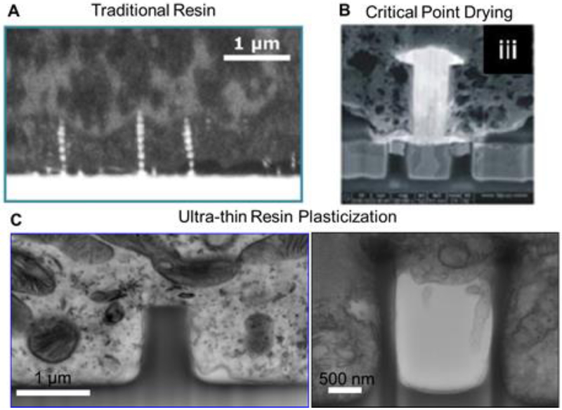
- Belu A, Schnitker J, Bertazzo S, Neumann E, Mayer D, Offenhäusser A, and Santoro F. 2016. “Ultra-Thin Resin Embedding Method for Scanning Electron Microscopy of Individual Cells on High and Low Aspect Ratio 3D Nanostructures.” Journal of Microscopy 263 (1): 78–86. - PubMed
Publication types
MeSH terms
Substances
Grants and funding
LinkOut - more resources
Full Text Sources
Other Literature Sources
