. 2018 Sep;32(9):4744-4752.
doi: 10.1096/fj.201701474RR. Epub 2018 Mar 23.

Deletion of choline acetyltransferase in enteric neurons results in postnatal intestinal dysmotility and dysbiosis

Affiliations

Deletion of choline acetyltransferase in enteric neurons results in postnatal intestinal dysmotility and dysbiosis

Carl D Johnson et al. FASEB J. 2018 Sep.

Abstract

Acetylcholine (ACh)-synthesizing neurons are major components of the enteric nervous system (ENS). They release ACh and peptidergic neurotransmitters onto enteric neurons and muscle. However, pharmacological interrogation has proven inadequate to demonstrate an essential role for ACh. Our objective was to determine whether elimination of ACh synthesis during embryogenesis alters prenatal viability, intestinal function, the neurotransmitter complement, and the microbiome. Conditional deletion of choline acetyltransferase ( ChAT), the ACh synthetic enzyme, in neural crest-derived neurons ( ChAT-Null) was performed. Survival, ChAT activity, gut motility, and the microbiome were studied. ChAT was conditionally deleted in ENS neural crest-derived cells. Despite ChAT absence, mice were born live and survived the first 2 wk. They failed to gain significant weight in the third postnatal week, dying between postnatal d 18 and 30. Small intestinal transit of carmine red was 50% slower in ChAT-Nulls vs. WT and ChAT- Het. The colons of many neonatal ChAT-Null mice contained compacted feces, suggesting dysmotility. Microbiome analysis revealed dysbiosis in ChAT-Null mice. Developmental deletion of ChAT activity in enteric neurons results in proximal gastrointestinal tract dysmotility, critically diminished colonic transit, failure to thrive, intestinal dysbiosis, and death. ACh is necessary for sustained gut motility and survival of neonatal mice after weaning.-Johnson, C. D., Barlow-Anacker, A. J., Pierre, J. F., Touw, K., Erickson, C. S., Furness, J. B., Epstein, M. L., Gosain, A. Deletion of choline acetyltransferase in enteric neurons results in postnatal intestinal dysmotility and dysbiosis.

Keywords: acetylcholine; development; enteric nervous system; microbiome.


Conflict of interest statement

The authors thank Brian Torres for assistance in performing assays, Dr. Timur Mavlyutov (University of Wisconsin-Madison) for assistance with confocal imaging, and Dr. June Dahl (University of Wisconsin-Madison) for suggestions on the manuscript. This work was supported by U.S. National Institutes of Health, National Institute of Diabetes and Digestive and Kidney Diseases Grants K08DK098271, R03DK114543, and P30DK42086; by an American Pediatric Surgical Association Foundation Scholars Award (to A.G.); and by an American College of Surgeons George H.A. Clowes Career Development Award (to A.G.). The authors declare no conflicts of interest.

Figures

Figure 1
Figure 1
ChAT activity is absent in the GI tract of ChAT-Null mice. ChAT activity in extracts of P22–P26 WT, ChAT-Het, and ChAT-Null gut. Assays were performed in triplicate. Blank controls (absence of sample), absence of Ac-CoA (No Ac-CoA), and total counts made in the absence of choline kinase were performed in duplicate. Incubation times were 1 h for spinal cord samples and 2 h for intestinal samples. Fc values of [3H]choline to acetyl-[3H]choline corrected for substrate depletion were calculated and recorded (n = 2–5 preparations/genotype). Error bars represent sem. *P < 0.05.
Figure 2
Figure 2
Loss of ChAT results in reduced weight over the first 3 wk of life. Weights of P21 WT, ChAT-Het, and ChAT-Null mice. Means ± sem of WT (n = 9), ChAT-Het (n = 9), and ChAT-Null (n = 7) animals measured during the first 3 wk of life. Error bars represent sem. *P < 0.001.
Figure 3
Figure 3
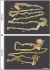
ChAT-Null mice have colonic fecal impaction. Bright field image of representative ChAT-Het and ChAT-Null GI tracts dissected from P4 animals. Note the reduced length of the intestine in the ChAT-Null compared with the ChAT-Het. Fecal material is compacted in the cecum and colon of ChAT-Null mice. Scale bar, 1 cm.
Figure 4
Figure 4
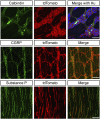
Expression of calbindin, CGRP, and substance P is preserved in ChAT-Null mice. Upper row: calbindin-IR neurons and neuronal processes (green) are present within the myenteric ganglia, which are endogenously labeled with tdTomato (red, center panel) of a P25 ChAT-Null cecum. Right panel shows a merged image with the neuronal marker, Hu-IR, (blue). Scale bar, 50 µm. Middle row: CGRP-IR processes (green) are abundant within ChAT-Null small intestine. Center panel shows tdTomato+ neurons; a merged image can be seen in the right panel. CGRP-IR processes project between myenteric ganglia. Scale bar, 100 µm. Bottom row, left panel: substance P–IR processes (green) appear throughout the smooth muscle of a P25 ChAT-Null colon. Most substance P–IR processes also express tdTomato, as shown in the merged right panel. Scale bar, 100 µm.
Figure 5
Figure 5
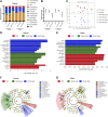
Dysbiosis in the gut of ChAT-Null mice. 16S rRNA sequencing of bacterial communities in WT, ChAT-Het, and ChAT-Null cecum and colon samples. A) Relative abundance of cecal and colonic bacterial phyla is shown for each genotype. B) Alpha diversity was measured by the Shannon Diversity Index, where ChAT-Null displayed lower diversity in both microbiota communities. C) Beta diversity dissimilarity was calculated with weighted UniFrac distances, where PC1 explained 33% of variability, and PC2 explained 20% of variability observed in cecal and colonic communities. D, E) To determine differentially represented taxa within each group, LDA LEfSe was calculated. Significantly differential taxa reaching logarithmic LDA scores of >2 are displayed for each group. Tenericutes were consistently represented by WT animals in both microbial communities, whereas Enterobacteriaceae were enriched in ChAT-Null samples. F, G) The differentially represented taxa determined by LEfSe were then graphed as cladograms to display phylogenic structure across taxonomic levels. Cladogram taxa abbreviated by letters are shown in the key in (F, G). These results demonstrate the shifted microbiota taxa were taxonomically related between genotypes, suggesting the altered luminal environment with deficient ChAT-directed specific shifts in the bacterial kingdom. Error bars represent sem.

References

    1. Qu Z. D., Thacker M., Castelucci P., Bagyánszki M., Epstein M. L., Furness J. B. (2008) Immunohistochemical analysis of neuron types in the mouse small intestine. Cell Tissue Res. 334, 147–161 10.1007/s00441-008-0684-7 - DOI - PubMed
    1. Erickson C. S., Lee S. J., Barlow-Anacker A. J., Druckenbrod N. R., Epstein M. L., Gosain A. (2014) Appearance of cholinergic myenteric neurons during enteric nervous system development: comparison of different ChAT fluorescent mouse reporter lines. Neurogastroenterol. Motil. 26, 874–884 10.1111/nmo.12343 - DOI - PMC - PubMed
    1. Sang Q., Young H. M. (1996) Chemical coding of neurons in the myenteric plexus and external muscle of the small and large intestine of the mouse. Cell Tissue Res. 284, 39–53 10.1007/s004410050565 - DOI - PubMed
    1. Furness J. B. (2012) The enteric nervous system and neurogastroenterology. Nat. Rev. Gastroenterol. Hepatol. 9, 286–294 10.1038/nrgastro.2012.32 - DOI - PubMed
    1. Bornstein J. C., Costa M., Grider J. R. (2004) Enteric motor and interneuronal circuits controlling motility. Neurogastroenterol. Motil. 16(Suppl 1), 34–38 10.1111/j.1743-3150.2004.00472.x - DOI - PubMed

Publication types

LinkOut - more resources