Review
. 2018 Mar 23;19(4):958.
doi: 10.3390/ijms19040958.

Micro-Economics of Apoptosis in Cancer: ncRNAs Modulation of BCL-2 Family Members

Affiliations
Review

Micro-Economics of Apoptosis in Cancer: ncRNAs Modulation of BCL-2 Family Members

Lidia Villanova et al. Int J Mol Sci. .

Abstract

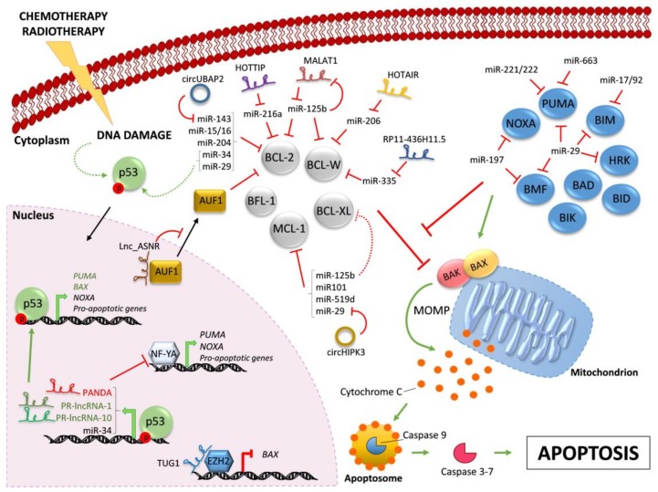
In the last few years, non-coding RNAs (ncRNAs) have been a hot topic in cancer research. Many ncRNAs were found to regulate the apoptotic process and to play a role in tumor cell resistance to treatment. The apoptotic program is on the frontline as self-defense from cancer onset, and evasion of apoptosis has been classified as one of the hallmarks of cancer responsible for therapy failure. The B-cell lymphoma 2 (BCL-2) family members are key players in the regulation of apoptosis and mediate the activation of the mitochondrial death machinery in response to radiation, chemotherapeutic agents and many targeted therapeutics. The balance between the pro-survival and the pro-apoptotic BCL-2 proteins is strictly controlled by ncRNAs. Here, we highlight the most common mechanisms exerted by microRNAs, long non-coding RNAs and circular RNAs on the main mediators of the intrinsic apoptotic cascade with particular focus on their significance in cancer biology.

Keywords: BCL-2 family; apoptosis; cancer; long non-coding RNAs; microRNAs; therapy resistance.


Conflict of interest statement

The authors declare no conflict of interest.

Figures

Figure 1
Figure 1
Regulation of BCL-2 family members by ncRNAs. Chemotherapy and/or radiotherapy induce DNA damage that leads to the activation of p53 (dashed curve green arrow). Active p53 translocates into the nucleus and drives the transcription (bold green arrows) of the indicated pro-apoptotic proteins and ncRNAs. Indicated lncRNAs control the transcription of pro-apoptotic proteins by different mechanisms. The pro-apoptotic multi-BH domain BAX and BAK are kept inactive by the interaction with pro-survival BCL-2-like proteins (in grey). All BH3-only proteins (in blue) can indirectly activate BAX/BAK by sequestering their pro-survival BCL-2-like relatives (red line), whereas only some of them can also trigger BAX/BAK activation through direct binding (green arrow). Active BAX/BAK oligomerize on the mitochondria membrane and lead to mitochondrial outer membrane permeabilization (MOMP) and consequent Cytochrome C release in the cytoplasm. Activation of downstream caspase cascade ultimately triggers apoptosis. In the cytoplasm, microRNAs negatively regulate BH3-only proteins and pro-survival BCL-2 family members. Indicated lncRNAs and circRNAs act as miR-sponges, counteracting miR-mediated regulation of pro-survival proteins. Further, MALAT1 is down-regulated by miR-125b, in a negative feedback-loop. Red lines indicate inhibition; green arrows indicate activation. Continuous arrows and lines refer to direct regulation; dashed arrows and lines indicate indirect regulation. Black arrows indicate nuclear/cytoplasmic translocations.

Similar articles

Cited by

References

    1. Kerr J.F., Wyllie A.H., Currie A.R. Apoptosis: A basic biological phenomenon with wide-ranging implications in tissue kinetics. Br. J. Cancer. 1972;26:239–257. doi: 10.1038/bjc.1972.33. - DOI - PMC - PubMed
    1. Adams J.M., Cory S. The Bcl-2 apoptotic switch in cancer development and therapy. Oncogene. 2007;26:1324–1337. doi: 10.1038/sj.onc.1210220. - DOI - PMC - PubMed
    1. Fulda S., Galluzzi L., Kroemer G. Targeting mitochondria for cancer therapy. Nat. Rev. Drug Discov. 2010;9:447–464. doi: 10.1038/nrd3137. - DOI - PubMed
    1. Willis S.N., Chen L., Dewson G., Wei A., Naik E., Fletcher J.I., Adams J.M., Huang D.C. Proapoptotic Bak is sequestered by Mcl-1 and Bcl-xL, but not Bcl-2, until displaced by BH3-only proteins. Genes Dev. 2005;19:1294–1305. doi: 10.1101/gad.1304105. - DOI - PMC - PubMed
    1. Willis S.N., Fletcher J.I., Kaufmann T., van Delft M.F., Chen L., Czabotar P.E., Ierino H., Lee E.F., Fairlie W.D., Bouillet P., et al. Apoptosis initiated when BH3 ligands engage multiple Bcl-2 homologs, not Bax or Bak. Science. 2007;315:856–859. doi: 10.1126/science.1133289. - DOI - PubMed

LinkOut - more resources