Immunomolecular Characterization of MIC-1, a Novel Antigen in Babesia bigemina, Which Contains Conserved and Immunodominant B-Cell Epitopes that Induce Neutralizing Antibodies
- PMID: 29570654
- PMCID: PMC6024600
- DOI: 10.3390/vetsci5020032
Immunomolecular Characterization of MIC-1, a Novel Antigen in Babesia bigemina, Which Contains Conserved and Immunodominant B-Cell Epitopes that Induce Neutralizing Antibodies
Abstract
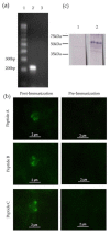
Babesia bigemina is one of the most prevalent species causing bovine babesiosis around the world. Antigens involved in host cell invasion are vaccine targets for this disease but are largely unknown in this species. The invasion process of Babesia spp. into erythrocytes involves membrane proteins from the apical complex. A protein stored in the micronemes, called Micronemal Protein 1 (MIC-1), contains a sialic acid binding domain that participates in the invasion process of host cells and is a vaccine candidate in other apicomplexan parasites. It is not known if there is a homologous gene for mic-1 in B. bigemina. Therefore, the aim of this study was to characterize the mic-1 gene homologue in Babesia bigemina. A gene was found with a microneme adhesive repeat (MAR) domain in the predicted amino acid sequence. Transcription was determined by reverse transcription polymerase chain reaction (RT-PCR). Subsequently, antibodies against peptides containing conserved B-cell epitopes were used to confirm the expression of MIC-1 in intraerythrocytic merozoites. The presence of anti MIC-1 antibodies in cattle naturally infected with B. bigemina was determined and up to 97.4% of the cattle sera (113 out of 116) identified MIC-1 using enzyme-linked immunosorbent assay (ELISA) methods. Finally, antibodies against MIC-1 were able to block 70% merozoite invasion in-vitro.
Keywords: Babesia bigemina; bovine babesiosis; micronemal proteins; sialic acid binding domain.
Conflict of interest statement
The authors declare no conflict of interest.
Figures


Similar articles
-
Identification and characterization of a Babesia bigemina thrombospondin-related superfamily member, TRAP-1: a novel antigen containing neutralizing epitopes involved in merozoite invasion.Parasit Vectors. 2020 Dec 1;13(1):602. doi: 10.1186/s13071-020-04469-5. Parasit Vectors. 2020. PMID: 33261638 Free PMC article.
-
RON2, a novel gene in Babesia bigemina, contains conserved, immunodominant B-cell epitopes that induce antibodies that block merozoite invasion.Parasitology. 2019 Nov;146(13):1646-1654. doi: 10.1017/S0031182019001161. Epub 2019 Sep 13. Parasitology. 2019. PMID: 31452491 Free PMC article.
-
Spherical Body Protein 4 from Babesia bigemina: A Novel Gene That Contains Conserved B-Cell Epitopes and Induces Cross-Reactive Neutralizing Antibodies in Babesia ovata.Pathogens. 2023 Mar 22;12(3):495. doi: 10.3390/pathogens12030495. Pathogens. 2023. PMID: 36986418 Free PMC article.
-
Genetic basis for GPI-anchor merozoite surface antigen polymorphism of Babesia and resulting antigenic diversity.Vet Parasitol. 2006 May 31;138(1-2):33-49. doi: 10.1016/j.vetpar.2006.01.038. Epub 2006 Mar 23. Vet Parasitol. 2006. PMID: 16551492 Review.
-
Designing blood-stage vaccines against Babesia bovis and B. bigemina.Parasitol Today. 1999 Jul;15(7):275-81. doi: 10.1016/s0169-4758(99)01471-4. Parasitol Today. 1999. PMID: 10377530 Review.
Cited by
-
Advances in Babesia Vaccine Development: An Overview.Pathogens. 2023 Feb 11;12(2):300. doi: 10.3390/pathogens12020300. Pathogens. 2023. PMID: 36839572 Free PMC article. Review.
-
Babesia bovis RON2 contains conserved B-cell epitopes that induce an invasion-blocking humoral immune response in immunized cattle.Parasit Vectors. 2018 Nov 3;11(1):575. doi: 10.1186/s13071-018-3164-2. Parasit Vectors. 2018. PMID: 30390674 Free PMC article.
-
The GP-45 Protein, a Highly Variable Antigen from Babesia bigemina, Contains Conserved B-Cell Epitopes in Geographically Distant Isolates.Pathogens. 2022 May 18;11(5):591. doi: 10.3390/pathogens11050591. Pathogens. 2022. PMID: 35631112 Free PMC article.
-
Recombinant Vaccinia Virus Expressing Plasmodium berghei Apical Membrane Antigen 1 or Microneme Protein Enhances Protection against P. berghei Infection in Mice.Trop Med Infect Dis. 2022 Nov 4;7(11):350. doi: 10.3390/tropicalmed7110350. Trop Med Infect Dis. 2022. PMID: 36355892 Free PMC article.
-
Report from the 'One Health' 9th Tick and Tick-Borne Pathogen Conference and the 1st Asia-Pacific Rickettsia Conference, Cairns, Australia, 27th August-1st September 2017.Vet Sci. 2018 Oct 2;5(4):85. doi: 10.3390/vetsci5040085. Vet Sci. 2018. PMID: 30279400 Free PMC article.
References
-
- Lew A., Jorgensen W. Molecular approaches to detect and study the organisms causing bovine tick borne diseases: babesiosis and anaplasmosis. Afr. J. Biotechnol. 2005;4:292–302. doi: 10.5897/AJB2005.000-3058. - DOI
LinkOut - more resources
Full Text Sources
Other Literature Sources
Research Materials
Miscellaneous

