Review
. 2018 May 11;293(19):7108-7116.
doi: 10.1074/jbc.R117.803239. Epub 2018 Mar 23.

Astrocytic glycogen metabolism in the healthy and diseased brain

Affiliations
Review

Astrocytic glycogen metabolism in the healthy and diseased brain

Lasse K Bak et al. J Biol Chem. .

Abstract

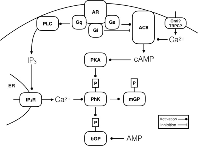
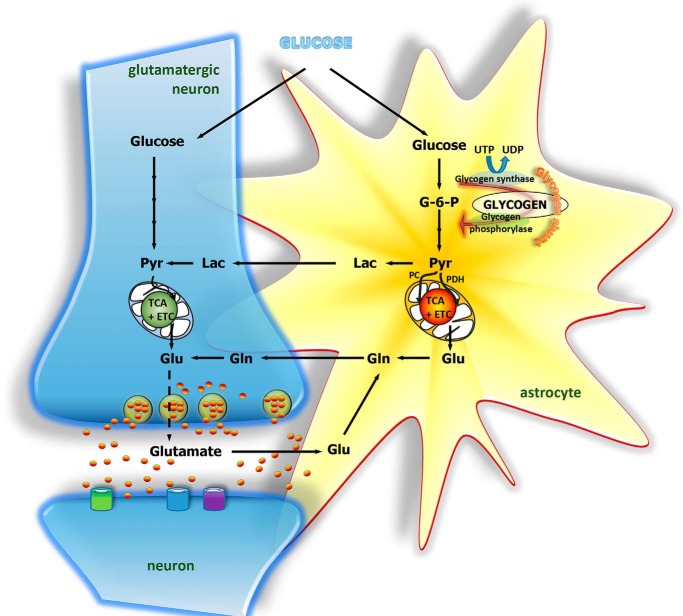
The brain contains a fairly low amount of glycogen, mostly located in astrocytes, a fact that has prompted the suggestion that glycogen does not have a significant physiological role in the brain. However, glycogen metabolism in astrocytes is essential for several key physiological processes and is adversely affected in disease. For instance, diminished ability to break down glycogen impinges on learning, and epilepsy, Alzheimer's disease, and type 2 diabetes are all associated with abnormal astrocyte glycogen metabolism. Glycogen metabolism supports astrocytic K+ and neurotransmitter glutamate uptake and subsequent glutamine synthesis-three fundamental steps in excitatory signaling at most brain synapses. Thus, there is abundant evidence for a key role of glycogen in brain function. Here, we summarize the physiological brain functions that depend on glycogen, discuss glycogen metabolism in disease, and investigate how glycogen breakdown is regulated at the cellular and molecular levels.

Keywords: astrocyte; brain; calcium; cyclic AMP (cAMP); disease; glycogen; neurological disease.


Conflict of interest statement

The authors declare that they have no conflicts of interest with the contents of this article

Figures

Figure 1.
Figure 1.
Cartoon depicting the two major signaling pathways regulating breakdown of glycogen. Glycogen phosphorylase brain (bGP) or muscle (mGP) forms are both activated by phosphorylation by a dedicated kinase, phosphorylase kinase (PhK). In addition, bGP is only fully active in the presence of ample levels of AMP. PhK, in turn, is activated by Ca2+ and phosphorylation by protein kinase A (PKA), and both signals are needed for full activation. In astrocytes, cAMP may be generated by plasma membrane-bound adenylate cyclase (AC), which in turn is regulated by the Gαs or Gαi protein-coupled adrenergic receptors (AR; see text for details). Depending on the isoform of AC expressed, Ca2+ flowing in via Orai or TRPC channels activated during store-operated Ca2+ entry may activate or inhibit the cAMP signal adding to the complexity; AC8 is activated by Ca2+ and is expressed in astrocytes. Finally, Gαq-coupled α1-adrenergic receptors may regulate glycogen breakdown via phospholipase C (PLC)-IP3 mediated release by IP3 receptors (IP3R) in the endoplasmic reticulum (ER).
Figure 2.
Figure 2.
Cartoon depicting glucose and glycogen metabolism in the brain as well as substrate transfer between astrocytes and neurons. In astrocytes, glucose may be metabolized via glycolysis or the glycogen shunt to pyruvate, which may be converted to lactate and transferred to neurons for oxidative metabolism to occur. Alternatively, pyruvate may enter the TCA cycle either by way of pyruvate dehydrogenase (PDH) or via pyruvate carboxylase (PC). Entrance of pyruvate via both of these pathways is required for de novo synthesis of glutamate and glutamine. Glutamine is not neuroactive and may be transferred to neurons to serve as precursor for glutamate synthesis. Following vesicular release of glutamate and interactions with receptors in the postsynaptic membrane, glutamate is cleared from the synapse mainly by transporters located in the astrocytic membrane. Glutamate can then be converted to glutamine and transferred to neurons, thereby completing the glutamate–glutamine cycle.

References

    1. Öz G., DiNuzzo M., Kumar A., Moheet A., and Seaquist E. R. (2015) Revisiting glycogen content in the human brain. Neurochem. Res. 40, 2473–2481 10.1007/s11064-015-1664-4 - DOI - PMC - PubMed
    1. Öz G., DiNuzzo M., Kumar A., Moheet A., Khowaja A., Kubisiak K., Eberly L. E., and Seaquist E. R. (2017) Cerebral glycogen in humans following acute and recurrent hypoglycemia: implications on a role in hypoglycemia unawareness. J. Cereb. Blood Flow Metab. 37, 2883–2893 10.1177/0271678X16678240 - DOI - PMC - PubMed
    1. Oz G., Kumar A., Rao J. P., Kodl C. T., Chow L., Eberly L. E., and Seaquist E. R. (2009) Human brain glycogen metabolism during and after hypoglycemia. Diabetes 58, 1978–1985 10.2337/db09-0226 - DOI - PMC - PubMed
    1. Duran J., Saez I., Gruart A., Guinovart J. J., and Delgado-García J. M. (2013) Impairment in long-term memory formation and learning-dependent synaptic plasticity in mice lacking glycogen synthase in the brain. J. Cereb. Blood Flow Metab. 33, 550–556 10.1038/jcbfm.2012.200 - DOI - PMC - PubMed
    1. Suzuki A., Stern S. A., Bozdagi O., Huntley G. W., Walker R. H., Magistretti P. J., and Alberini C. M. (2011) Astrocyte-neuron lactate transport is required for long-term memory formation. Cell 144, 810–823 10.1016/j.cell.2011.02.018 - DOI - PMC - PubMed

Publication types