. 2018 Mar 23;8(1):5136.
doi: 10.1038/s41598-018-22985-4.

Altered Insulin/Insulin-Like Growth Factor Signaling in a Comorbid Rat model of Ischemia and β-Amyloid Toxicity

Affiliations

Altered Insulin/Insulin-Like Growth Factor Signaling in a Comorbid Rat model of Ischemia and β-Amyloid Toxicity

Zareen Amtul et al. Sci Rep. .

Abstract

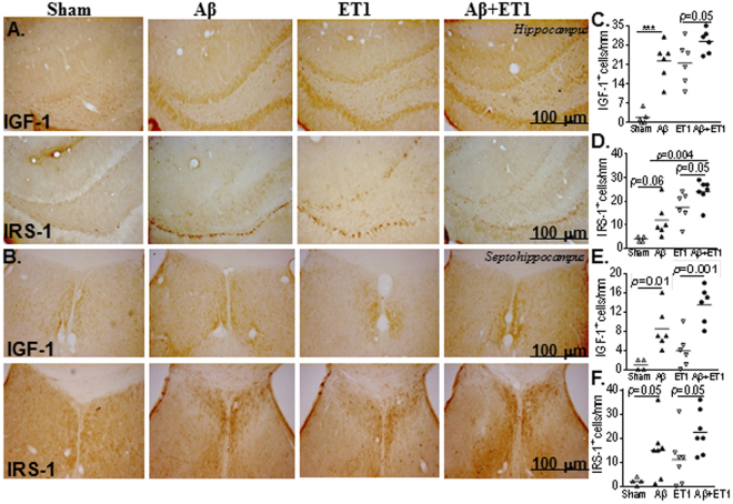
Ischemic stroke and diabetes are vascular risk factors for the development of impaired memory such as dementia and/or Alzheimer's disease. Clinical studies have demonstrated that minor striatal ischemic lesions in combination with β-amyloid (Aβ) load are critical in generating cognitive deficits. These cognitive deficits are likely to be associated with impaired insulin signaling. In this study, we examined the histological presence of insulin-like growth factor-I (IGF-1) and insulin receptor substrate (IRS-1) in anatomically distinct brain circuits compared with morphological brain damage in a co-morbid rat model of striatal ischemia (ET1) and Aβ toxicity. The results demonstrated a rapid increase in the presence of IGF-1 and IRS-1 immunoreactive cells in Aβ + ET1 rats, mainly in the ipsilateral striatum and distant regions with synaptic links to the striatal lesion. These regions included subcortical white matter, motor cortex, thalamus, dentate gyrus, septohippocampal nucleus, periventricular region and horizontal diagonal band of Broca in the basal forebrain. The alteration in IGF-1 and IRS-1 presence induced by ET1 or Aβ rats alone was not severe enough to affect the entire brain circuit. Understanding the causal or etiologic interaction between insulin and IGF signaling and co-morbidity after ischemia and Aβ toxicity will help design more effective therapeutics.


Conflict of interest statement

The authors declare no competing interests.

Figures

Figure 1
Figure 1
Striatum and thalamus: Low resolution images at bregma levels +0.48 mm show cerebral injury in the striatal lesion core of ET1 and Aβ + ET1 rats. (A) The dotted rectangles indicate the region of the high magnification images in B,C,D and E. High resolution immunostaining indicates staining for striatal expression of APP fragments including β-amyloid (B) and FJB degeneration (C) as well as IGF-1 and IRS-1 in the ipsilateral striatum (D) and thalamus (E) of sham, Aβ, ET1 and Aβ+ET1 rats Plots show quantitative assessment of β-amyloid (F), FJB (G), IGF-1 and IRS-1 staining in the striatum (H and I) and thalamus (J and K) of sham, Aβ, ET1 and Aβ+ET1 rats, respectively. (B,C,F and G are courtesy from5).
Figure 2
Figure 2
Cortex and subcortical white matter: High resolution immunostaining indicates staining for IGF-1 and IRS-1 in the ipsilateral cortex (A) and subcortical white matter (B) of sham, Aβ, ET1 and Aβ+ET1 rats. Plots show quantitative assessment of IGF-1 and IRS-1 staining in the cortex (C and D) and subcortical white matter (E and F) of sham, Aβ, ET1 and Aβ + ET1 rats.
Figure 3
Figure 3
Hippocampus and septohippocampus: High resolution immunostaining indicates staining for IGF-1 and IRS-1 in the ipsilateral dentate gyrus of hippocampus (A) and the septohippocampus (B) of sham, Aβ, ET1 and Aβ + ET1 rats. Plots show quantitative assessment of IGF-1 and IRS-1 staining in the dentate gyrus (C and D) and septohippocampus (E and F) of sham, Aβ, ET1 and Aβ + ET1 rats, *p < 0.05, **p < 0.01.
Figure 4
Figure 4
Periventricles and basal forebrain: High resolution immunostaining indicates staining for IGF-1 and IRS-1 in the ipsilateral periventricles (A) and horizontal diagonal band (HDB) of Broca in basal forebrain (B) of sham, Aβ, ET1 and Aβ + ET1 rats. Plots show quantitative assessment of IGF-1 and IRS-1 staining in the periventricles (C and D) and HDB (E and F) of sham, Aβ, ET1 and Aβ + ET1 rats.

References

    1. Hachinski VC, B. J. Vascular dementia. Neurology. 1993;43:2159–60. doi: 10.1212/WNL.43.10.2159-a. - DOI - PubMed
    1. Snowdon DA. Brain Infarction and the Clinical Expression of Alzheimer Disease.The Nun Study. JAMA: The Journal of the American Medical Association. 1997;277:813. doi: 10.1001/jama.1997.03540340047031. - DOI - PubMed
    1. Thiel A, et al. The temporal dynamics of poststroke neuroinflammation: a longitudinal diffusion tensor imaging-guided PET study with 11C-PK11195 in acute subcortical stroke. J. Nucl. Med. 2010;51:1404–12. doi: 10.2967/jnumed.110.076612. - DOI - PubMed
    1. Yang Y, Estrada EY, Thompson JF, Liu W, Rosenberg GA. Matrix Metalloproteinase-Mediated Disruption of Tight Junction Proteins in Cerebral Vessels is Reversed by Synthetic Matrix Metalloproteinase Inhibitor in Focal Ischemia in Rat. Journal of Cerebral Blood Flow & Metabolism. 2007;27:697–709. doi: 10.1038/sj.jcbfm.9600375. - DOI - PubMed
    1. Amtul Z, et al. Comorbid rat model of ischemia and β-Amyloid toxicity: Striatal and cortical degeneration. Brain Pathology. 2015;25:24–32. doi: 10.1111/bpa.12149. - DOI - PMC - PubMed

Publication types

MeSH terms