

Accurate label-free 3-part leukocyte recognition with single cell lens-free imaging flow cytometry
- PMID: 29573668
- PMCID: PMC5933530
- DOI: 10.1016/j.compbiomed.2018.03.008
Accurate label-free 3-part leukocyte recognition with single cell lens-free imaging flow cytometry
Abstract
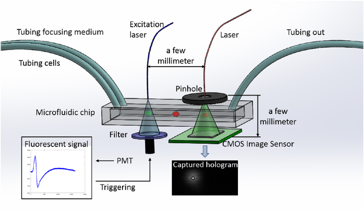
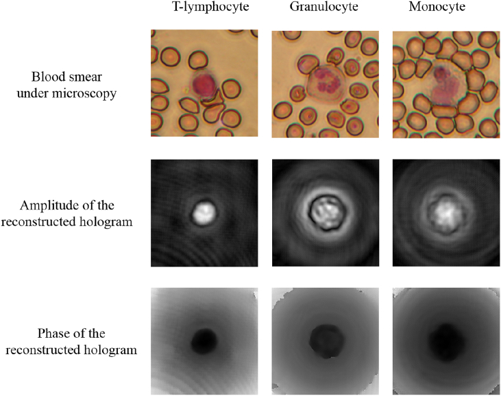
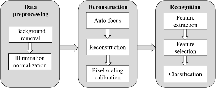
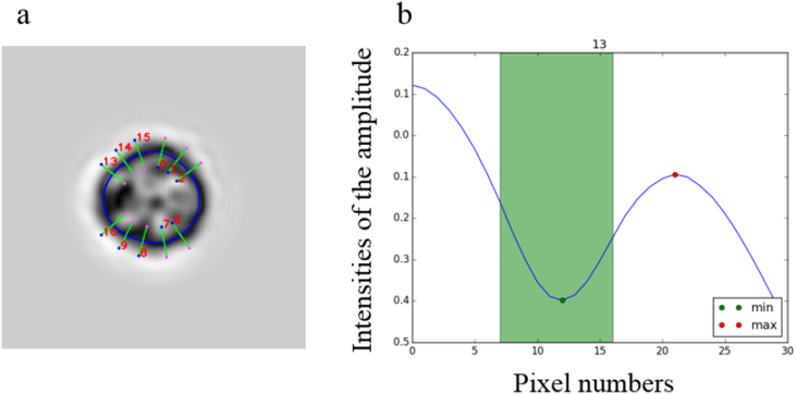
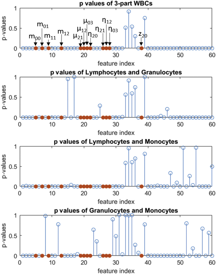
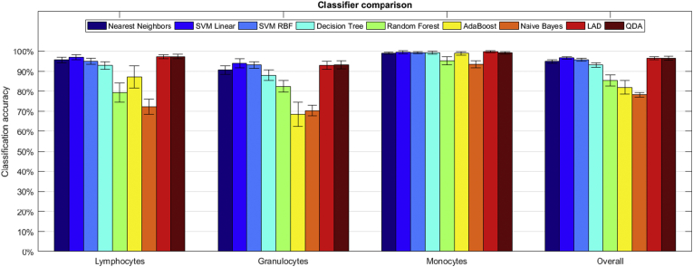
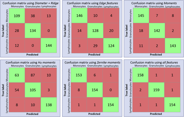
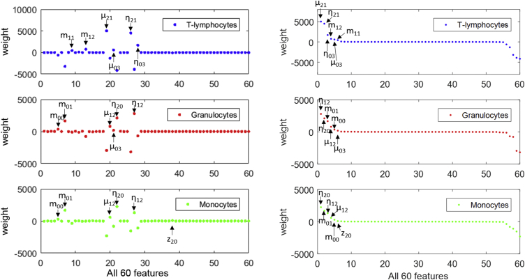
Three-part white blood cell differentials which are key to routine blood workups are typically performed in centralized laboratories on conventional hematology analyzers operated by highly trained staff. With the trend of developing miniaturized blood analysis tool for point-of-need in order to accelerate turnaround times and move routine blood testing away from centralized facilities on the rise, our group has developed a highly miniaturized holographic imaging system for generating lens-free images of white blood cells in suspension. Analysis and classification of its output data, constitutes the final crucial step ensuring appropriate accuracy of the system. In this work, we implement reference holographic images of single white blood cells in suspension, in order to establish an accurate ground truth to increase classification accuracy. We also automate the entire workflow for analyzing the output and demonstrate clear improvement in the accuracy of the 3-part classification. High-dimensional optical and morphological features are extracted from reconstructed digital holograms of single cells using the ground-truth images and advanced machine learning algorithms are investigated and implemented to obtain 99% classification accuracy. Representative features of the three white blood cell subtypes are selected and give comparable results, with a focus on rapid cell recognition and decreased computational cost.
Keywords: Flow cytometry; Hologram; Lens-free imaging; Three-part differential; White blood cell.
Copyright © 2018 The Authors. Published by Elsevier Ltd.. All rights reserved.
Figures
Similar articles
-
Single Cell Analysis of Stored Red Blood Cells Using Ultra-High Throughput Holographic Cytometry.Cells. 2021 Sep 17;10(9):2455. doi: 10.3390/cells10092455. Cells. 2021. PMID: 34572104 Free PMC article.
-
Three-part differential of unlabeled leukocytes with a compact lens-free imaging flow cytometer.Lab Chip. 2015 Feb 21;15(4):1123-32. doi: 10.1039/c4lc01131g. Lab Chip. 2015. PMID: 25537881
-
High accuracy label-free classification of single-cell kinetic states from holographic cytometry of human melanoma cells.Sci Rep. 2017 Sep 20;7(1):11943. doi: 10.1038/s41598-017-12165-1. Sci Rep. 2017. PMID: 28931937 Free PMC article.
-
Implementing machine learning methods for imaging flow cytometry.Microscopy (Oxf). 2020 Apr 8;69(2):61-68. doi: 10.1093/jmicro/dfaa005. Microscopy (Oxf). 2020. PMID: 32115658 Review.
-
Single-shot common-path off-axis digital holography: applications in bioimaging and optical metrology [Invited].Appl Opt. 2021 Feb 1;60(4):A195-A204. doi: 10.1364/AO.404208. Appl Opt. 2021. PMID: 33690370 Review.
Cited by
-
On the use of deep learning for phase recovery.Light Sci Appl. 2024 Jan 1;13(1):4. doi: 10.1038/s41377-023-01340-x. Light Sci Appl. 2024. PMID: 38161203 Free PMC article. Review.
-
Label-Free Identification of White Blood Cells Using Machine Learning.Cytometry A. 2019 Aug;95(8):836-842. doi: 10.1002/cyto.a.23794. Epub 2019 May 13. Cytometry A. 2019. PMID: 31081599 Free PMC article.
-
Deep-Learning Based Label-Free Classification of Activated and Inactivated Neutrophils for Rapid Immune State Monitoring.Sensors (Basel). 2021 Jan 13;21(2):512. doi: 10.3390/s21020512. Sensors (Basel). 2021. PMID: 33450866 Free PMC article.
-
Machine learning issues and opportunities in ultrafast particle classification for label-free microflow cytometry.Sci Rep. 2020 Nov 26;10(1):20724. doi: 10.1038/s41598-020-77765-w. Sci Rep. 2020. PMID: 33244129 Free PMC article.
-
Label-Free High-Throughput Leukemia Detection by Holographic Microscopy.Adv Sci (Weinh). 2018 Oct 11;5(12):1800761. doi: 10.1002/advs.201800761. eCollection 2018 Dec. Adv Sci (Weinh). 2018. PMID: 30581697 Free PMC article.
References
-
- Maton D., Hopkins J., McLaughlin C.W., Johnson S., Warner M.Q., LaHart D., Wright J.D., Kulkarni D.V. Englewood Cliffs; New Jersey, US: 1997. Human Biology and Health. ISBN 0-13-981176-1.
-
- Heikali D., Di Carlo D. A niche for microfluidics in portable hematology analyzers. JALA: J. Assoc. Lab. Autom. 2010;15(4):319–328.
-
- Shapiro H.M., Perlmutter N.G. Personal cytometers: slow flow or no flow? Cytometry. 2006;69(7):620–630. - PubMed
-
- Eisenstein M. Cell sorting: divide and conquer. Nature. 2006;441(7097):1179–1185. - PubMed
-
- Li Q., Wang Y., Liu H., He X., Xu D., Wang J., Guo F. Leukocyte cells identification and quantitative morphometry based on molecular hyperspectral imaging technology. Comput. Med. Imag. Graph. 2014;38(3):171–178. - PubMed
Publication types
MeSH terms
LinkOut - more resources
Full Text Sources
Other Literature Sources
