

Computational neuroanatomy of baby brains: A review
- PMID: 29574033
- PMCID: PMC6150852
- DOI: 10.1016/j.neuroimage.2018.03.042
Computational neuroanatomy of baby brains: A review
Abstract
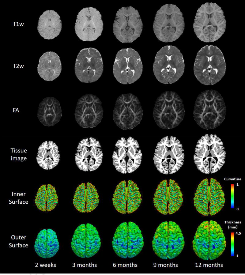
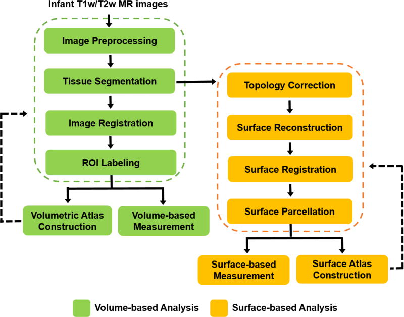
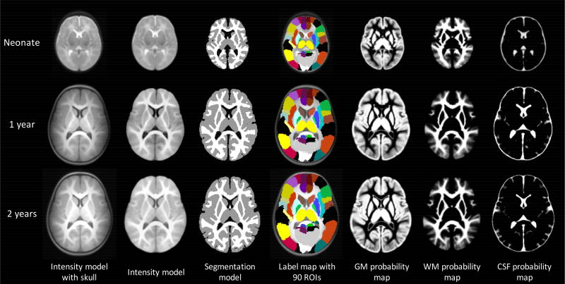
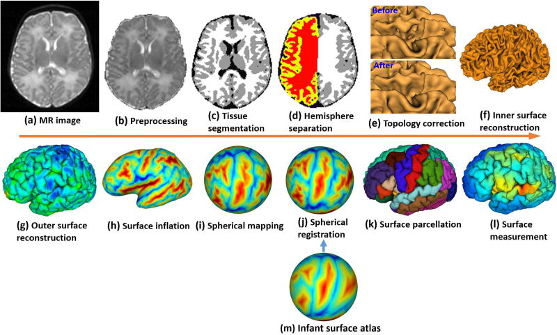
The first postnatal years are an exceptionally dynamic and critical period of structural, functional and connectivity development of the human brain. The increasing availability of non-invasive infant brain MR images provides unprecedented opportunities for accurate and reliable charting of dynamic early brain developmental trajectories in understanding normative and aberrant growth. However, infant brain MR images typically exhibit reduced tissue contrast (especially around 6 months of age), large within-tissue intensity variations, and regionally-heterogeneous, dynamic changes, in comparison with adult brain MR images. Consequently, the existing computational tools developed typically for adult brains are not suitable for infant brain MR image processing. To address these challenges, many infant-tailored computational methods have been proposed for computational neuroanatomy of infant brains. In this review paper, we provide a comprehensive review of the state-of-the-art computational methods for infant brain MRI processing and analysis, which have advanced our understanding of early postnatal brain development. We also summarize publically available infant-dedicated resources, including MRI datasets, computational tools, grand challenges, and brain atlases. Finally, we discuss the limitations in current research and suggest potential future research directions.
Keywords: Brain atlas; Cortical surface; Infant brain; Parcellation; Registration; Segmentation.
Copyright © 2018 Elsevier Inc. All rights reserved.
Figures
References
-
- Akazawa K, Chang L, Yamakawa R, Hayama S, Buchthal S, Alicata D, Andres T, Castillo D, Oishi K, Skranes J, Ernst T, Oishi K. Probabilistic maps of the white matter tracts with known associated functions on the neonatal brain atlas: Application to evaluate longitudinal developmental trajectories in term-born and preterm-born infants. NeuroImage. 2016;128:167–179. - PMC - PubMed
-
- Alexander B, Murray AL, Loh WY, Matthews LG, Adamson C, Beare R, Chen J, Kelly CE, Rees S, Warfield SK. A new neonatal cortical and subcortical brain atlas: the Melbourne Children’s Regional Infant Brain (M-CRIB) atlas. NeuroImage. 2017;147:841–851. - PubMed
-
- Aljabar P, Heckemann RA, Hammers A, Hajnal JV, Rueckert D. Multi-atlas based segmentation of brain images: Atlas selection and its effect on accuracy. NeuroImage. 2009;46:726–738. - PubMed
Publication types
MeSH terms
Grants and funding
LinkOut - more resources
Full Text Sources
Other Literature Sources
