. 2018 Mar 24;7(7):e007394.
doi: 10.1161/JAHA.117.007394.

Modeling Short QT Syndrome Using Human-Induced Pluripotent Stem Cell-Derived Cardiomyocytes

Affiliations

Modeling Short QT Syndrome Using Human-Induced Pluripotent Stem Cell-Derived Cardiomyocytes

Ibrahim El-Battrawy et al. J Am Heart Assoc. .

Abstract

Background: Short QT syndrome (SQTS), a disorder associated with characteristic ECG QT-segment abbreviation, predisposes affected patients to sudden cardiac death. Despite some progress in assessing the organ-level pathophysiology and genetic changes of the disorder, the understanding of the human cellular phenotype and discovering of an optimal therapy has lagged because of a lack of appropriate human cellular models of the disorder. The objective of this study was to establish a cellular model of SQTS using human-induced pluripotent stem cell-derived cardiomyocytes (hiPSC-CMs).

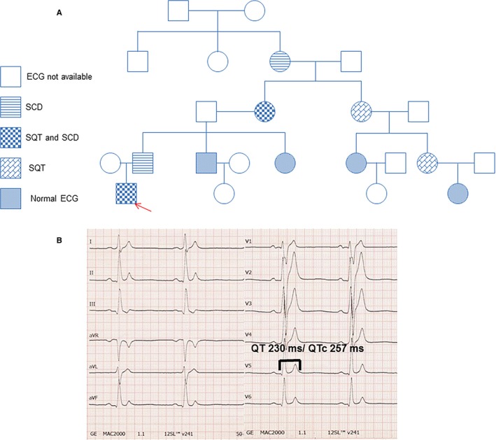
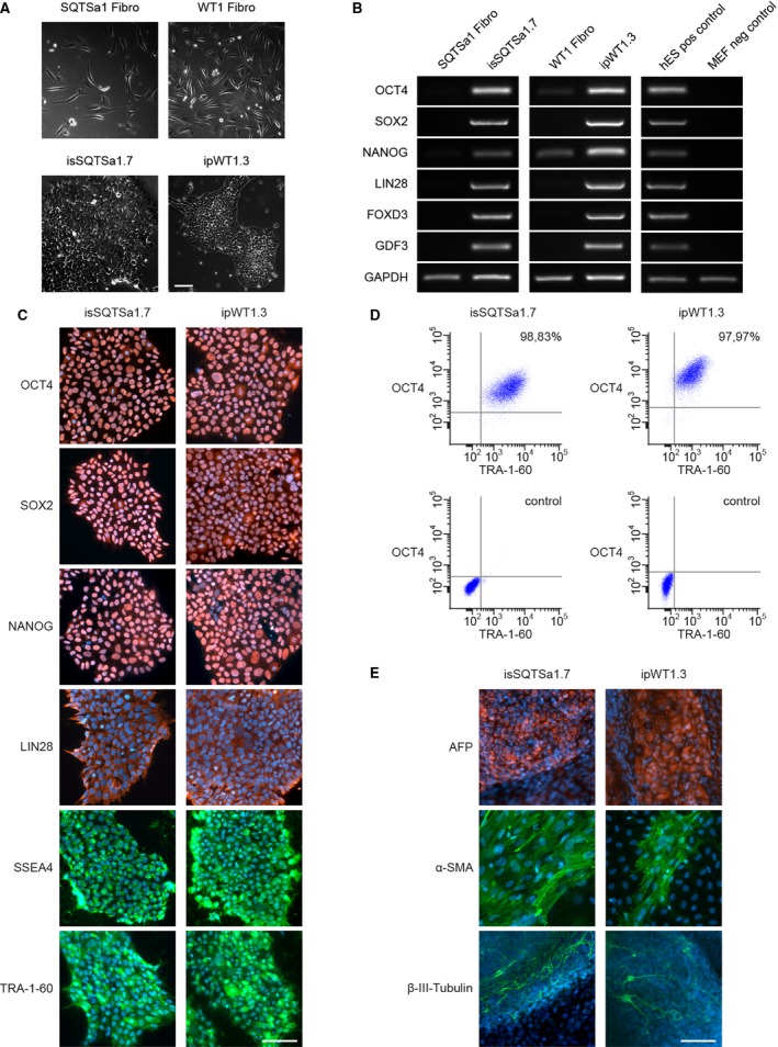
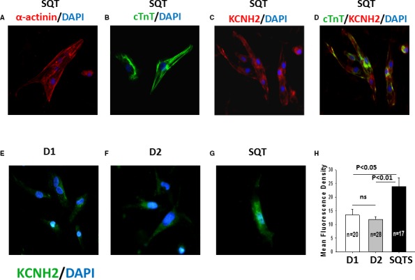
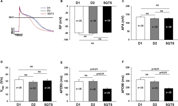
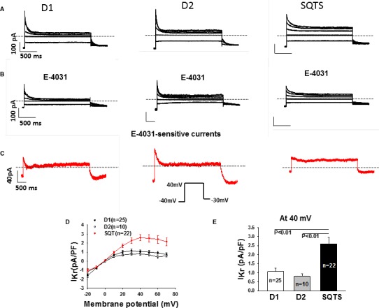
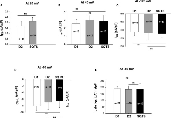
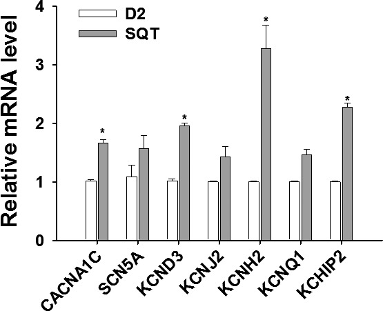
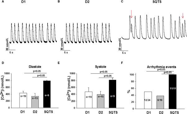
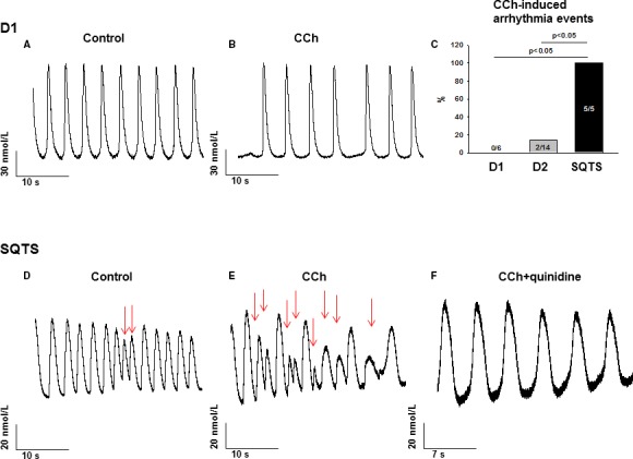
Methods and results: This study recruited 1 patient with short QT syndrome type 1 carrying a mutation (N588K) in KCNH2 as well as 2 healthy control subjects. We generated hiPSCs from their skin fibroblasts, and differentiated hiPSCs into cardiomyocytes (hiPSC-CMs) for physiological and pharmacological studies. The hiPSC-CMs from the patient showed increased rapidly activating delayed rectifier potassium channel current (IKr) density and shortened action potential duration compared with healthy control hiPSC-CMs. Furthermore, they demonstrated abnormal calcium transients and rhythmic activities. Carbachol increased the arrhythmic events in SQTS but not in control cells. Gene and protein expression profiling showed increased KCNH2 expression in SQTS cells. Quinidine but not sotalol or metoprolol prolonged the action potential duration and abolished arrhythmic activity induced by carbachol.

Conclusions: Patient-specific hiPSC-CMs are able to recapitulate single-cell phenotype features of SQTS and provide novel opportunities to further elucidate the cellular disease mechanism and test drug effects.

Keywords: arrhythmia (heart rhythm disorders); arrhythmia (mechanisms); ion channel; short QT syndrome.


Figures

Figure 1
Figure 1
A, The pedigree of the family with SQTS. The patient recruited for this study is marked by the arrow. B, The ECG from the SQTS patient shows a classic SQTSECG pattern. SCD indicates sudden cardiac death; SQT, short QT syndrome (SQTS).
Figure 2
Figure 2
Pluripotent characteristics of human induced pluripotent stem cell (hiPSC) lines. A, The hiPSC lines of short QT syndrome (SQTS) (SQTSa1.7) and donor 2 (D2) (ipWT1.3) generated from skin fibroblasts (upper panel) display a typical morphology for human pluripotent stem cells (lower panel). B, In comparison to donor's fibroblasts, generated iPSC lines show expression of endogenous pluripotency markers SOX2 (sex determining region Y‐ box 2), OCT4 (octamer‐binding transcription factor 4), NANOG (pron. nanOg, homeobox protein), LIN28 (lin‐28 homolog A), FOXD3 (Forkhead Box D3), and GDF3 (growth differentiation factor‐3) at mRNA level proven by reverse transcription‐polymerase chain reaction. Human embryonic stem cells (hESCs) were used as positive control, and mouse embryonic fibroblasts (MEFs) were used as negative control. C, Generated hiPSC lines express pluripotency markers OCT4, SOX2, NANOG, LIN28, SSEA4 (stage‐specific embryonic antigen 4), and TRA‐1‐60 as shown by immunofluorescence staining. Nuclei are co‐stained with DAPI (4′, 6‐diamidino‐2‐phenylindole). D, Flow cytometry analysis of pluripotency markers OCT4 and TRA‐1‐60 reveals a homogeneous population of pluripotent cells in generated iPSC lines. E, Spontaneous differentiation potential of generated hiPSC lines was analyzed by embryoid body formation and germ‐layer specific marker expression. Immunocytochemical staining of spontaneously differentiated hiPSC lines shows expression of endodermal marker AFP (α‐fetoprotein), mesodermal‐specific α‐SMA (α‐smooth muscle actin), and ectodermal βIII‐tubulin. Nuclei are co‐stained with DAPI. Scale bars: 100 μm.
Figure 3
Figure 3
KCNH2 channels are upregulated in SQTS (short QT syndrome)‐cardiomyocytes. Shown are the immunostaining for detecting cardiomyocyte markers (SQTS; A and B) and KCNH2 (rapidly activating delayed rectifier potassium channel) proteins located in the whole cell (SQTS; C and D) and the cell membrane of hiPSCCMs (human induced pluripotent stem cell–derived cardiomyocytes) from the healthy donors (D1, D2; E and F) and the patient (SQTS; G). Nuclear staining was induced with DAPI (4′, 6‐diamidino‐2‐phenylindole) (blue). A, Red: Fluorescein isothiocyanate (FITC)‐conjugated α‐actinin antibody at day 40 after differentiation (α‐actinin). B, Green: FITC‐conjugated cTnT2 (human cardiac troponin T) antibody at day 40 after differentiation (cTnT). C, Red: Human KCNH2 antibody at day 40 after differentiation. D, Green: FITC ‐conjugated cTnT2 antibody, red: human KCNH2 antibody at day 40 after differentiation. Nuclear staining was induced with DAPI (blue). E through G, Green: Human KCNH2 antibody located in the cell membrane at day 40 after differentiation. H, The mean fluorescence density of hiPSCCMs from the donors (D1, D2) and the patient (SQTS). ns indicates not significant difference.
Figure 4
Figure 4
Shortened action potential duration (APD) in SQTS (short QT syndrome)‐cardiomyocytes. A, Representative traces of action potentials (AP) in control‐ (D1 and D2) and SQTS‐cardiomyocytes (SQTS). B, Mean values of resting potentials (RP). C, Mean values of action potential amplitude (APA). D, Mean values of maximal upstroke velocity of AP (Vmax). E, Mean values of APD at 50% repolarization (APD50). F, Mean values of APD at 90% repolarization (APD90). Values given are mean±SEM. n, number of cells. ns indicates not significant difference.
Figure 5
Figure 5
Enhanced IKr in SQTS (short QT syndrome)‐cardiomyocytes. Ikr (rapidly activating delayed rectifier potassium channel) was recorded D1, D2, and SQTS‐cells depolarized from −20 to +70 mV with a holding potential of −40 mV to measure the steady‐state currents. The pulses were repolarized to −30 mV to check the tail currents. Symmetrical Cs+ concentrations (140/140 mmol/L) were used for the current recordings. E‐4031 (3 μmol/L) was applied to isolate IKr. A and B, Representative families of IKr traces (at −20, 0, 20, 40, and 60 mV) in a D1, D2, and SQTS‐cell in absence (A) and presence (B) of E‐4031. C, Representative traces of E‐4031 sensitive currents (IKr). D, Comparison of I–V (current–voltage relationship) curves of Ikr from D1, D2, and SQTS‐cells. E, Comparison of IKr density at 40 mV between donor and the patient. Values given are mean±SEM. n, number of cells.
Figure 6
Figure 6
Changes in ion channel currents in SQTS (short QT syndrome)‐cardiomyocytes. Ion channel currents were recorded in the control‐ (D1, D2) and SQTS‐cells (SQTS). A, Mean values of IK s (slowly activating delayed rectifier potassium channel). B, Mean values of peak Ito (transient outward potassium channel current). C, Mean values of IK 1 (inward rectifier potassium channel current). D, Mean values of peak IC aL (L‐type calcium channel current). E, Mean values of the area under curve of TTX (tetrodotoxin) sensitive late IN a (sodium channel current). Values given are mean±SEM; n, number of cells; ns (not significant), P>0.05.
Figure 7
Figure 7
Changes in mRNA expression of ion channels in SQTS (short QT syndrome)‐cardiomyocytes. The relative mRNA levels of each ion channel were analyzed by qPCR (quantitative PCR) in hiPSCCMs (human induced pluripotent stem cell–derived cardiomyocytes) of the patient (STQS) and compared with control cells (D2). Values given are mean±SEM (from 3 biological replicates). *P<0.05 vs control (D2).
Figure 8
Figure 8
Increased intracellular Ca2+ level and arrhythmic events in SQTS (short QT syndrome)‐cardiomyocytes. A and B, Representative Ca2+ transients in donor (D1, D2) cells. C, Representative Ca2+ transients in a SQTS‐human induced pluripotent stem cell–derived cardiomyocytes with early afterdepolarization‐ and delayed afterdepolarization‐like triggered activities (marked by red arrows). D, Mean values of diastolic Ca2+ concentration. E, Mean values of systolic Ca2+ concentration. F, Percentages of cells showing arrhythmic (triggered) events. Values given are mean±SEM; n, number of cells; ns (not significant), P>0.05.
Figure 9
Figure 9
Carbachol increased and quinidine abolished arrhythmic events in SQTS (short QT syndrome)‐cardiomyocytes. A and B, Representative Ca2+ transients in a donor (D1) cell in absence (A) and presence (B) of 10 μmol/L carbachol (CCh). C, Percentages of cells showing CCh‐induced arrhythmia events. D and E, Representative Ca2+ transients in a SQTS‐cell in absence (D) and presence (E) of 10 μmol/L CCh. The red arrows indicate the arrhythmia events. F, Representative Ca2+ transients in a SQTS‐cell in presence of 10 μmol/L CCh plus 10 μmol/L quinidine.

References

    1. Algra A, Tijssen JG, Roelandt JR, Pool J, Lubsen J. Qt interval variables from 24 hour electrocardiography and the two year risk of sudden death. Br Heart J. 1993;70:43–48. - PMC - PubMed
    1. Guerrier K, Kwiatkowski D, Czosek RJ, Spar DS, Anderson JB, Knilans TK. Short QT interval prevalence and clinical outcomes in a pediatric population. Circ Arrhythm Electrophysiol. 2015;8:1460–1464. - PubMed
    1. Priori SG, Blomstrom‐Lundqvist C, Mazzanti A, Blom N, Borggrefe M, Camm J, Elliott PM, Fitzsimons D, Hatala R, Hindricks G, Kirchhof P, Kjeldsen K, Kuck KH, Hernandez‐Madrid A, Nikolaou N, Norekval TM, Spaulding C, Van Veldhuisen DJ. 2015 ESC Guidelines for the management of patients with ventricular arrhythmias and the prevention of sudden cardiac death: the Task Force for the Management of Patients with Ventricular Arrhythmias and the Prevention of Sudden Cardiac Death of the European Society of Cardiology (ESC). Endorsed by: Association for European Paediatric and Congenital Cardiology (AEPC). Eur Heart J. 2015;36:2793–2867. - PubMed
    1. Giustetto C, Di Monte F, Wolpert C, Borggrefe M, Schimpf R, Sbragia P, Leone G, Maury P, Anttonen O, Haissaguerre M, Gaita F. Short QT syndrome: clinical findings and diagnostic‐therapeutic implications. Eur Heart J. 2006;27:2440–2447. - PubMed
    1. Gaita F, Giustetto C, Bianchi F, Wolpert C, Schimpf R, Riccardi R, Grossi S, Richiardi E, Borggrefe M. Short QT syndrome: a familial cause of sudden death. Circulation. 2003;108:965–970. - PubMed

Publication types

MeSH terms

Supplementary concepts