Review
. 2018 Aug;53(8):893-906.
doi: 10.1007/s00535-018-1451-5. Epub 2018 Mar 24.

To die or not to die: death signaling in nonalcoholic fatty liver disease

Affiliations
Review

To die or not to die: death signaling in nonalcoholic fatty liver disease

Yuko Akazawa et al. J Gastroenterol. 2018 Aug.

Abstract

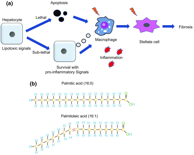
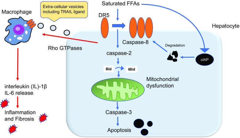
Non-alcoholic fatty liver disease (NAFLD) is an emerging liver disease worldwide. In subset of patients, NAFLD progresses to its advanced form, nonalcoholic steatohepatitis (NASH), which is accompanied with inflammation and fibrosis. Saturated free fatty acid-induced hepatocyte apoptosis is a feature of NASH. Death signaling in NASH does not always result in apoptosis, but can alternatively lead to the survival of cells presenting signs of pro-inflammatory and pro-fibrotic signals. With the current lack of established treatments for NASH, it is important to understand the molecular mechanisms responsible for disease development and progression. This review focuses on the latest findings in hepatocyte death signaling and discusses possible targets for intervention, including caspases, death receptor and c-Jun N-terminal kinase 1 signaling, oxidative stress, and endoplasmic reticulum stress, as well as epigenomic factors.

Keywords: Apoptosis; Endoplasmic reticulum stress; Free fatty acids; Non-alcoholic fatty liver disease; Nonalcoholic steatohepatitis.


Figures

Fig. 1
Fig. 1
a Lethal and sublethal signaling during lipotoxity. Lethal lipotoxic signals induced by free fatty acids induce hepatocyte apoptosis, which are engulfed by macrophage, initiating inflammatory and fibrotic reactions. When the apoptotic signaling pathways are initiated by the apoptosis in not executed, sublethal lipotoxic signals release vesicles that are delivered to macrophages, potentially participating in progression of NASH by elevating inflammation. b. Simplified image of saturated free fatty acid (palmitic acid) and unsaturated free fatty acid (palmitoleic acid). Palmitoleic acid has a double bond within the carbon chain, such that when it is incorporated in the double membrane, it confers better fluidity to the membrane. Palmitic acid does not have double bonds in its carbon chain, making the membrane more rigid and less fluid
Fig. 2
Fig. 2
Integrated model of death receptor (DR)-mediated apoptosis and inflammation in non-alcoholic steatohepatitis (NASH). Free fatty acids (FFAs) induce aggregation of DR5 on the cell membrane and activate caspase following the formation of a complex with DR5. Caspase 8 activation results in cleavage of BH3-only protein Bid to truncated (t)-Bid, thereby contributing to mitochondrial dysfunction and cell death. Degradation of cIAP, an anti-apoptotic protein, also contributes to lipoapoptosis. Saturated FFA-induced DR5 activation also causes the release of extracellular vesicles in a Rho-GTPase-dependent manner. TNF-related apoptosis-inducing ligand (TRAIL)-containing vesicles are recognized by DR5 on macrophages, eliciting an inflammatory response and fibrosis
Fig. 3
Fig. 3
Lipotoxicity in hepatocytes and cholangiocytes. In hepatocytes, mixed lineage kinase 3-glycogen synthase kinase 3β (MLK-3-GSK)-activated c-Jun N-terminal kinase (JNK) phosphorylation plays a central role in apoptosis. JNK and endoplasmic reticulum (ER) stress protein CHOP cooperatively induce the BH3-only protein PUMA, leading to apoptosis. JNK binds to Sab, an mitochondrial outer membrane protein, which promotes translocation of JNK to mitochondria, inducing reactive oxygen species (ROS). MiR-34a also contributes to lipoapoptosis. Although saturated free fatty acids (FFAs) are poorly incorporated into lipid droplets, steatosis occurs to some extent in hepatocytes. In contrast, FFAs do not induce steatosis in cholangiocytes. Lipotoxicity is mediated by forkhead box O3 (Foxo3)-stimulated miR-34a and PUMA up-regulation. JNK does not contribute to cell death in cholangiocytes
Fig. 4
Fig. 4
Endoplasmic reticulum (ER) stress-mediated apoptosis. Saturated free fatty acids (FFAs) induce ER stress, activating protein kinase R-like ER kinase (PERK), inositol requiring 1 (IRE1), and activating transcription factor 6 (ATF6). These signals first activate the protective unfolded protein response (UPR); however, excessive stress leads to activation of apoptotic signaling, mainly through PERK-mediated CHOP up-regulation. miR-615-3p, which suppresses CHOP expression, is decreased during FFA treatment. Augmentation of miR-615-3p levels partially inhibits CHOP expression and cell death induced by FFA-mediated ER stress. Maresin 1 (MAR1) resolves lipotoxicity and ER stress by up-regulating UPR pro-survival mechanisms and preventing the excessive stimulation of pro-apoptotic pathways. MAR1 is also able to attenuate ER stress in macrophages, restoring Kupffer cell phagocytic capacity to clear apoptotic hepatocytes
Fig. 5
Fig. 5
Interactions between lethal and nonlethal pro-inflammatory signaling by saturated free fatty acids (FFAs) and potential interventions for NASH. Saturated FFAs induce apoptotic signaling through multiple pathways, including endoplasmic reticulum (ER) stress, death receptor (DRS) and c-Jun N-terminal kinase (JNK) signaling, non-coding RNAs, reactive oxygen species (ROS), and dysregulation of autophagy. These signals ultimately merge to induce mitochondrial dysfunction and the release of the executioner caspase 3, leading to cell death. Sublethal amounts of FFAs induce pro-inflammatory signaling in parenchymal cells, leading to inflammation and fibrosis. Based on recent discoveries regarding pathways involved in FFA-induced toxicity, the proteins and drugs highlighted in green are potential interventional targets for NASH

References

    1. Williams CD, Stengel J, Asike MI, et al. Prevalence of nonalcoholic fatty liver disease and nonalcoholic steatohepatitis among a largely middle-aged population utilizing ultrasound and liver biopsy: a prospective study. Gastroenterology. 2011;140(1):124–131. - PubMed
    1. Seki Y, Kakizaki S, Horiguchi N, et al. Prevalence of nonalcoholic steatohepatitis in Japanese patients with morbid obesity undergoing bariatric surgery. J Gastroenterol. 2016;51(3):281–289. - PubMed
    1. Watanabe S, Hashimoto E, Ikejima K, et al. Evidence-based clinical practice guidelines for nonalcoholic fatty liver disease/nonalcoholic steatohepatitis. J Gastroenterol. 2015;50(4):364–377. - PubMed
    1. Yan J, Xie W, Ou WN, et al. Epidemiological survey and risk factor analysis of fatty liver disease of adult residents, Beijing, China. J Gastroenterol Hepatol. 2013;28(10):1654–1659. - PubMed
    1. Tokushige K, Hashimoto E, Kodama K. Hepatocarcinogenesis in non-alcoholic fatty liver disease in Japan. J Gastroenterol Hepatol. 2013;28(Suppl 4):88–92. - PubMed